What is Causing My Heel Pain?

Lateral X-ray of a Calcaneum demonstrating a spur.

Image via Wikipedia

Heel pain can be caused by a number of factors.  One of the most common is a heel spur, medically referred to as a calcaneal spur.

A heel spur is an outgrowth of bone in the shape of a small spire.  If it’s at the bottom or side of the heel bone (calcaneous), it can be painful to walk.  It is thought that heel spurs form as a result of stress on a tendinous insertion into to the heel, but some heel spurs aren’t associated with the area where a tendon inserts into the heel.

Whenever offshoots of bone form (bone spurs), it is due to a history of stress to the bone, either gradual formation following a single impact trauma such as a car accident; or cumulative impact forces over time, such as from sports.  For the foot bones, an example of the latter would be years of playing basketball or ballet dancing.  The repetitive pounding on the joints of the foot can cause bone cells at the impact area to secrete more bone matrix as a compensatory response.  In the spine and large weight bearing joints (knee, hip) they are called osteophytes.

In some cases, the heel spur stops producing pain; perhaps due to resorbption or even callous formation neutralizing its ability to press against tissue. For those that don’t, surgery can be done by a podiatrist or orthopedic surgeon to smooth down the heel spur.

TREATMENT:

In the meantime, avoiding impact trauma to the heel is important (running, jumping).  You can buy special shoe inserts that help reduce pressure on the heel spur.  Comfortable shoes are a must as well; make sure they are wide and have a thick rubber, shock absorbing sole.  The Z-Coil shoe brand incorporates a large spring at the heel to minimize shock/ impact forces to the foot when walking.

Sitting in a chair and rolling a cold beer or soda can (unopened) under your foot can help stretch the foot and ease some of the pain.  A golf ball can provide more focused massage to the bottom of the foot.  Press downward with enough pressure to get a good stretch and massage, but not so much to increase the heel pain.

Dorsal and plantar aspects of foot

Image via Wikipedia

Lastly, consider exercising your feet by walking barefoot around the house and outside (walking barefoot indoors is not as effective due to the flatness of the floor; bumpy and uneven is better).  Walking in shoes actually weaken the foot over time by preventing the intrinsic foot muscles and joints from exercising and experiencing their full range of motion.  You can also consider getting the Vibram foot glove.  This interesting shoe fits on your foot like a glove, allowing some independent movement of the toes.

Plantar fascitis is a condition where the plantar fascia (a wide, band-like ligament connecting the heel to the forefoot) pulls off a thin layer of periosteum (membrane lining the bone) from the heel and results in inflammation and pain at the front, bottom part of the heel.  It can make walking very uncomfortable.

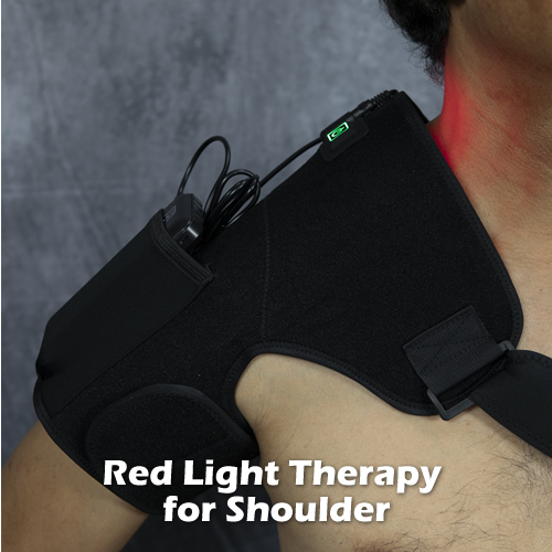
The best approach to treating plantar fascitis is to rest your foot for a few days:  no running, no walking up hill; walk only on flat surfaces.  Use Red Light Therapy and Pulsed EMF for 15 minutes, twice a day for 2-3 weeks to accelerate healing of tissues, and for reducing any inflammation that is causing pain.  Red light therapy uses photobiomodulation to accelerate cell metabolism; thus accelerating healing of tissues.

Compression fractures and hairline (stress) fractures can also cause heel pain.  If you think you’ve fracture your heelbone, have it X-rayed.   You’ll likely get a foot cast, avoid impact to the heel for several months, and wait until the fracture heals (no pun intended).

If your heel bone fractures easily, it could be due to a bone cysts.  See your doctor on solutions for dealing with bone cysts.

Carpal Tunnel Syndrome

A rigid splint can keep the wrist straight.

Image via Wikipedia

Carpal tunnel syndrome is a condition where one of the major nerves of the arm gets compressed in the wrist.  It can lead to pain, numbness, and tingling in the hands.  Advanced symptoms are muscle weakness in the hands, muscle atrophy (shrinking), especially of the thumb pad;  and loss of motor coordination in fine dexterity skills, like buttoning a blouse.

The three main nerves that are responsible for controlling the arm are the ulnar, median, and radial nerves.  The median nerve, like its name implies, travels down the middle of the arm.  It passes through the carpal tunnel  which is just above the crease in the wrist before splitting into branches that go to the thumb, index, middle, and inner half of the ring finger.

The carpal tunnel is a small diameter hole formed by the wrist bones and the transverse carpal ligament.  It contains the tendons that flex the fingers (flexor tendons), and the median nerve.  Pressure as light as a penny can adversely affect nerve tissue, so any pressure increase in the carpal tunnel will over time injure the median nerve.

The most common cause of increased pressure in the carpal tunnel is thickening of the flexor tendons due to long term repetitive use of the fingers such as in typing.  Over time the tendons press the median nerve against the rigid transverse carpal ligament.  The nerve loses oxygen and it starts to malfunction.  Left alone, the damage will be permanent as nerves have a limited ability to regenerate.

Other possible causes are prior injury to the wrist that narrows the carpal tunnel and arthritic or other pathological changes in the wrist bones that cause them to occlude the tunnel.

Pregnancy and thyroid conditions may mimic symptoms of carpal tunnel syndrome.

TREATMENT:

Scars from carpal tunnel release surgery. Two ...

Image via Wikipedia

If the symptoms are advanced (pain, numbness, tingling especially at NIGHT and loss of hand coordination and muscle atrophy) see your doctor.  The doctor should refer you to a hand specialist who may order a nerve conduction test to diagnose carpal tunnel syndrome.  If your test is positive, you may be referred for physical therapy, which will involve mostly stretching and hand exercises.  If that doesn’t work, you may be offered a cortisone injection and exercise prescription, a wrist brace and orders to avoid prolonged hand usage.  The last option is carpal tunnel release surgery, where the transverse carpal ligament is surgically cut to relieve pressure in the tunnel.

If your condition is not advanced, do the following:

If your job or hobby requires lots of finger and hand activity, there is a good chance that this is the cause of your symptoms.  Check your work station set up and ensure the following:

a.  Keyboard should be low enough so that your fingers are at the level of the keyboard when:

  • your upper arms and shoulders are relaxed; your upper arms (above the elbow) are to the side of your body almost touching;
  • your elbows are bent 90-100 degrees
  • your wrists are straight or even bent slightly downward

The most important part is having your shoulders relaxed.  To see if you are doing it right, using your right hand press the top of your left upper shoulder, from the neck down to the shoulder joint.  It should be relatively soft.  If it is not, you are unconsciously contracting the neck and upper trapezius muscles and lifting the arm.

If you find you can’t accomplish the above, due to your desk being too high, you need to get an adjustable keyboard tray and install it under your desk.  Place the keyboard on this tray and lower and angle the tray so that you can meet these requirements (see video below on how to do this).

b.  The top 1/3 of your monitor screen should be at eye level.  Use phone books or a monitor lift to get it to this position.   Place the monitor close enough that you don’t  have to bend your neck forward to see text on your screen, or adjust your screen settings to magnify the text.

c. The mouse and frequently used equipment should be close so that you don’t have to reach forward for them.  Your keyboard tray should have an attached mouse pad; use it.

d. Remember to keep your head in a position where your ears are directly over your shoulders.

e. Every few minutes, relax your hands and wrists for 20-30 seconds.

f. Every hour do the wrist, neck and shoulder exercises in the video.

g. When symptoms are gone, you can do wrist strengthening exercises.

Exercise for Thoracic Outlet Syndrome

The thoracic outlet is an “opening” at the base of the neck, on both sides, where the nerves and artery that service the arm on that side exit through as they descend into the arm.  It is formed by the anterior and medial scalene muscles, the first rib, and the clavicle (collar bone).

If the thoracic outlet narrows in any way, due to one or more of these structures, neurovascular compression is possible (compression of the nerves and artery).  This can result in shoulder pain, neck pain, weakness in the affected arm and hand, numbness and tingling in the arm down to the hands, especially the last three fingers; and in some cases swelling of the arm.  Carrying something like a heavy bag on the affected shoulder makes the symptoms worse.

In thoracic outlet syndrome there is usually a history of prior injury/trauma to the shoulder girdle, such as a car accident or sports injury.  People with long necks and long arms are said to be more predisposed to this condition as the weight of the arm and length of the neck tend to put more stress in the thoracic outlet area.

Left alone without resolution, it is possible to develop permanent nerve damage resulting in chronic pain, weakness in the arm and hand, and reduced sensation in the inner arm area.

What to Do if You Suspect You Have Thoracic Outlet Syndrome

If you suspect you have thoracic outlet syndrome, see your doctor or an experienced chiropractor.  There are some simple orthopedic tests that can be done to see if you likely have TOS.  An MRI study can help visualize the thoracic outlet and identify if there is swelling; and needle electromyograph (EMG) can determine if the arm muscles are getting the proper amount of nerve flow.

If the diagnosis is confirmed, physical therapy exercises are usually prescribed to help open the thoracic outlet space.   In some cases, imaging studies show a fibrous band  responsible for compressing the neurovascular structure; in these cases surgery may be an option.  This involves the scalene muscles — three, short muscles that connect the lower neck vertebrae to the ribcage.

In the meantime, try doing this exercise.  It is designed to create more space between the collar bone and the ribcage.  If this is where your compression is occurring, it may help.


How to Stop Tension Headaches

Headaches come in many different forms; too many to include in one post.

The causation can be neurological, vascular, mechanical, chemical and even psychosomatic.  Diagnosis can be challenging, as most headaches have the common symptom of, well, head ache.  The factors that vary include duration, location of pain (back of head, front of head, one side of head), pain pattern (constant, pulsating, repeating), and accompanying symptoms (dizziness, nausea, sensitivity to light and sound, auras).

This post is about tension headaches, perhaps the most common type.

Symptoms include constant, pressure like pain often described as a tightening band around the head.  The muscles of the back of the neck and tops of the shoulders are usually hypertonic (tense and taught).   Pain is felt behind the eyes.  Tension headaches can be mild to the point where the person goes on about his day until it wears off; or they can be intense and incapacitating, causing the person to take aspirin or Tylenol.

It is generally believed that tension headaches can be triggered by stress, dehydration, working in front of a bright computer monitor for extended periods; looking at a screen (TV, computer, movie screen) that has constantly moving images with changing light; and engaging in heavy mental tasks (studying, calculating numbers, reading conceptually-complex material like law cases, etc.).

More esoteric causes are previous trauma that affected the neck, like a car crash, and environmental stimuli (pollen, mites, carpet fumes, atomized copier toner, exposure to hazardous chemicals).

In my experience, people who have a history of severe whiplash from a car accident are more likely to have recurring tension headaches.  Even if the accident was ten or more years ago.

Whiplash is the violent, alternating extension and flexion of the neck due to a short but powerful impact force or short acceleration-deceleration.  Low impact car accidents and a jerky roller coaster ride are common examples.

The accident can leave the cervical (neck) vertebrae out of proper position relative to adjacent vertebrae, and change the dynamics of neck movement.  Nerves that regulate muscle contraction in the neck and and back of head can get injured or stretched as a result, and can cause the muscles to stiffen during certain times.

TREATMENT:

If you are engaging in heavy mental activities, give yourself a couple of hours break.  Turn off the TV; stay away from the computer and all screens for that matter.  Basically, you want to shut off excessive visual stimulation.

Seek silence and solace.  Find a nice park,  go for a nature hike.  Another option– meditate in a dark room; concentrate on deep breathing and  relaxing the muscles in the back of your neck and throughout your body.  Drink water throughout the day.  No coffee or cigarettes; they are stimulants.  No alcohol.

Place an ice pack on your forehead (put kitchen towelette on your forehead to prevent ice burn), OR one under your neck with a cervical roll supporting it (DON’T do both, the coldness may be too much stimuli).

If you have a history of a whiplash car accident, and you get tension headaches quite regularly, there’s a very good chance you have misaligned cervical vertebrae affecting your cord and/or nerve roots.   Probably a “reversed” curve, which looks like a “kink” or sudden angle change on a side-view neck x-ray. You will want to do exercises to stretch the neck and get it back to a lordotic curved shape.

Use a neck roll to bend your neck into a lordotic (reverse C- shape) curve while lying on your back on the floor.  Simply touch the floor with the back of your head ten times by arching your neck over the roll.  Then, turn and stretch your neck to the left and hold for 2 seconds; then to the right and hold for 2 seconds; 10 times to each side.  Do 3-4 times throughout the day.

You may also consider getting evaluated by an experienced chiropractor, and definitely getting a neck x-ray to visualize the shape of your cervical spine.   Adjustments, exercises, and lordotic traction can help bring your neck into proper alignment, and reduce pressure to your nerves, saving you from those annoying headaches.

Lastly, consider using red light and pulsed EMF to eliminate tension headaches.  This is a good investment if you have recurring headaches.

Red light therapy is using 630 nm wavelength light to reduce pain and inflammation.  Light at this wavelength gets absorbed by cell structures and basically increases circulation, vasodilates blood vessels and dampens inflammation.  Some tension headaches are vascular in nature, so this should help reduce symptoms.

Pulsed EMF is the application of weak electromagnetic fields to the body, to provide extra energy for cells needing it.  It tends to improve cell membrane transport of nutrients and waste, and improve molecular transport including red blood cell mobility.

Watch this video I made that explains how to do it:

What Happens When You “Pull” Your Leg Muscle?

The Achilles' tendon. PD image from Gray's Ana...

Image via Wikipedia

If there’s one thing that can stop you dead  in your tracks, it’s a pulled calf muscle.  The word “pulled muscle” usually refers to an involuntary, painful spasm that occurs without a single, forceful impact which characterizes a typical muscle sprain/ strain.

You’ll feel your calf muscle contract by itself, and sometimes oscillate (twitch) reflexively causing you to quickly bend your knee to stop the progressing contraction.  A deep, painful sensation is felt at the myofascial junction that eventually forms the Achilles tendon.  If you’re lucky, sometimes you can prevent it from spasming  if you immediately stop what you are doing at the first sign of the spasm (jerky, involuntary twitching) and focus on relaxing the leg.

Basically, pulled calf muscles occur after prolonged exercise of the legs, such as in long distance running, cycling, swimming, and uphill climbing.  Muscle contraction is mediated by a complex biological pathway that involves electrolytes, mainly calcium, phosphorus, and sodium ions.  These ions need to be available in order for the actin and myosin fibers to “ratchet” properly during contraction and relaxation.  Prolonged leg exertion, without replenishing lost electrolytes can lead to muscle spasms.  This is why sports teams use Gatorade to hydrate the athletes, and protect against muscle injuries.

Nocturnal (night) calf muscle spasms are a common occurrence in 2nd and 3rd trimester pregnant women.  It it thought to be caused by low electrolytes as a result of the pregnancy.  Eating foods rich in calcium and phosphorus can reduce these symptoms.

Lactic acid buildup in the myofascial sheath can also be a contributing factor to pulled muscles.  If more lactic acid is created that can be neutralized by the body, it can affect muscle contraction.

TREATMENT:

If the spasm got you before you could stop it, don’t panic.  The pain will work itself out in about 30 minutes.  If it is still present after an hour, see your doctor.

Drink an athletic sport drink to replenish your electrolytes.  If you have multivitamins at home, take 2 tablets with water.

Wrap an icepack around your calf, especially over the end of the muscle belly (that’s the start of the Achilles tendon); hold in place for 20 minutes.

Gradually, extend your knee; if you feel twitching again, stop and return to flexed knee position, ice applied; give it another 10 minutes.

When you can extend the knee fully without pain (all this is done sitting on the floor), gently test the calf muscle by bending your foot upwards toward your knee (called dorsiflexion).  Do this very slowly, and hold the stretch for10 seconds; repeat five times.

Next, attempt to walk.  That should be the end of your painful episode!  In the future, before you engage in arduous exercise involving a lot of legwork, load up on foods rich in electrolytes.  Most of those sports snacks include them, like Cliff Bars and others.  Try to get the healthy ones.

Receive a FREE, 30-Day Plan to Boost Your Health and Eliminate Pain!

Receive a FREE, 30-Day Plan to Boost Your Health and Eliminate Pain!

As a subscriber, you'll also learn the special methods used by experts in human biomechanics to fix body aches and pain the RIGHT way, long term. 

We'll also send you a Free eBook, Concepts of Self-Healing as a way of saying thanks.

Please check your email in 5 minutes to access your Special Report. Make sure to whitelist "newsletter@painandinjurydoctor.com" in your email client (Gmail, Yahoo, Outlook, etc.) so that you don't miss this valuable information. One way is to add this email to your email Contacts.