How I Got Trigger Finger and What I Did to Treat It

How I Got Trigger Finger and What I Did to Treat It

It’s been a while since my last article.  Between the weekly-changing COVID restrictions in my area and major house renovations, I have been delinquent with my life mission of helping others manage and heal their pain and injuries, on their own.  But today, I’m back on track.  Today, I’ll talk about a peculiar condition known as Trigger Finger. 

But first, a little background:

For those who ever engaged in do-it-yourself home renovations such as landscaping, fence building, paver-laying and bathroom and kitchen remodeling you know how much stress it can put on your body.  This is my story of how I developed trigger finger for the first time in my life, and serves as a “lessons learned” opportunity for others so that they can be spared the inconveniences of this condition..

For the last 10 years, I would categorize my daily physical activity as “moderate.”  I would go the gym and lift free weights (reps over max); do various cardio fitness classes such as the Les Mills classes and Bootcamp; and run 3 miles about 3x/week.  My average time in the gym I would say was 60-90 minutes, with about half of that actual exercising.  At home, I would be working on my website and producing videos.  This did require prolonged sitting, but I would get up every 30 minutes or so to walk around to relieve pressure to my lumbar spine.

Starting the second week of this past July, I started the aforementioned home renovation projects.  I basically went straight from moderate activity to short bursts of sustained intense activity, daily for over four weeks.  Since I didn’t have any major musculoskeletal impediments other than a chronic right AC (acromioclavicular) joint sprain, I moved freely as though I was in my 20s, which wasn’t such a good idea.  The combination of the intense movement patterns my body wasn’t used to, plus my age, took a significant toll after four weeks.

Here are some of the heavy labor activities that I engaged in:


  • Carrying heavy lumber from Home Depot and loading into a pickup truck, about 10 trips
  • Carrying 50 and 80 pound bags of concrete mix and sand, for my paver project, about 5 trips.
  • Used a 2-person auger (about 120 pounds; gasoline powered) to drill several 3’ deep post holes
  • Shoveled piles and piles of dirt (pickup truck loads—about 10x)
  • Hauled away bulk trash to the dumpster
  • Carried 100 clay 12”x12” paver squares (bricks) from a truck to my yard and positioned them carefully
  • Used hand tools that required hard gripping and/or twisting including various types of saws, wrenches and screwdrivers
  • Used vibrational tools including a miter saw, reciprocating saw, drill, and nail gun
risks for getting trigger finger

By the third week, I was starting to feel pain at my right AC joint, my left wrist, and both hands especially my right, dominant hand. Thankfully, despite frequent bending at the waist my lower back wasn’t affected.  I attributed the AC join pain to aggravation of the old strain (I rate it a Grade 2 or 3 sprain – partial tearing, but intact). What happened is the heavy lifting placed a repetitious load on that unstable joint, causing the acromion and distal clavicle to aggravate surrounding soft tissues, particularly the supraspinatous tendon, and the insertion points of the ligament.  My doctor suggested my pain was impingement syndrome—compression of the supraspinatous tendon where it passes below the acromion– which could be occurring, but I’m certain most of the pain is emanating from the joint itself because I can reproduce the pain simply by pressing it with my fingertip.  I’ll tell you how I’m treating this in the next article.

I believe my left wrist pain is a Grade 2 strain of the flexor ulnaris tendon where it inserts into the distal ulnar’s styloid process; caused when I lost control of the auger.  The auger is a very powerful machine that requires two people to operate (see picture above).  Not being familiar with using one, I wasn’t prepared for the powerful torque it generated, and lost control of it, hurting my wrist.

The third problem that I’m dealing with is trigger finger.  This is the first time I’ve had it and let me tell you, it’s not very pleasant.

Trigger finger is so named because as you attempt to straighten out your finger after closing your hand, the finger “catches” mid-way, and pain is felt in one or several joint capsules usually on the palmar side.  Then, as you power through the restriction the pain increases and a popping/snapping sensation occurs right before it straightens out, just like how a gun trigger offers gradual resistance then suddenly releases at a point.  See the short video below of my actual trigger finger taken this morning that explains this.

Trigger finger is a stenosing tenosynovitis disorder.  Stenosing means narrowing of a passageway in the body; tenosynovitis refers to inflammation of the tendon and synovium.  The synovium is a specialized layer of tissue surrounding the tendon in areas where it rubs against other structures in the body.  Synovium secretes synovial fluid, a biological lubricant that helps reduce friction where the tendon moves.  Synovium also lines the synovial joints of the body which include the hips, knees, shoulders, elbows, spine and joints of the hands and feet.


diagram of flexor tendons of the hand

There are three, main populations of trigger finger sufferers:  young children (up to 8 years old); trigger finger as a comorbidity to a primary disease; and adults experiencing trauma/ stress to the hands, typically in the 40s-50s.  It tends to affect women more, and the most common finger is the thumb although it can occur in any finger, and in multiple fingers at the same time.

In children, trigger finger is believed to be due to uneven growth rates of the hand flexor tendons and the ligaments, where the tendon growth outpaces the growth of the ligaments that hold them against the finger bones.

Trigger finger is observed to often occur alongside certain other diseases such as carpal tunnel syndrome, diabetes, hypothyroidism, gout, rheumatoid arthritis, and amyloidosis; each probably having different etiologies involving the dysfunction causing the primary disease.  Diabetics seem to be affected by trigger finger at a higher rate than the regular population, and it is uncertain why.  With diabetes mellitus, there are high levels of glucose in the blood, and usually high insulin levels.  Insulin is considered an anabolic hormone associated with tissue growth, so this may be a possible explanation for the increased incidence of trigger finger in diabetics, if the growth leads to tendon hypertrophy (enlargement).

For the third group, which the rest of this article will address, trigger finger is caused by hypertrophy and inflammation of the finger flexor tendons at the synovial sheath, typically from repetitious hand movements, especially those involving power gripping and vibration, making them chafe against the ligaments that hold them down to the finger bones (phalanges).  (Remember, ligaments connect two bones, while tendons connect a muscle to a bone; both are components of all moveable joints).  Imagine these ligaments as slips of Scotch tape forming a tunnel over the bone.  As the hypertrophied (enlarged) section of the tendon enters the narrow tunnel during extension (straightening out of the finger), it gets stuck in that tunnel momentarily; much like how a big person trying to climb out of a small bathroom window can get stuck before being able to make it through.  Then, as the tendon makes it past that entrance, it causes the popping sensation.

illustration of trigger finger locking

Orthopedic specialists identify the tendon-ligament structures involved in hand movement as pulleys.  Remember from basic physics, a pulley is one of the simple machines (the others being a lever, plane and gear).  This is an appropriate name because the tendons and ligaments accomplish work just like the cables and pulleys used in cranes.

pulley systems of the finger
Image courtesy of OrthoBullets.com

The A1 pulley is at the metacarpo-phalangeal joint, commonly called the knuckles.  It’s where the proximal phalanx connects to the respective metacarpal bone.  This is where trigger finger usually occurs.  Those who have it here feel the pain and popping/snapping on the palmar side of the knuckle.

The A2 pulley encircles the proximal phalanx, or first finger bone, from the knuckle.

The A3 pulley is at the PIP, or proximal interphalangeal joint—the first joint from the knuckle connecting the proximal and intermediate phalanges (first and second bones of the finger).  This is also a common area of trigger finger.

The A4 pulley encircles the intermediate phalanx (second bone of the finger from the knuckle).

The A5 pulley is at the DIP, or distal interphalangeal joint, the furthest joint of the finger connecting the intermediate and distal phalanges (second and third bones of the finger, from the knuckle).  Although triggering can happen here, it is less common.

Since the thumb is comprised of only two phalanges, it has an A1 and A2 pulley only.  Trigger finger affecting the thumb almost always occurs at the A1 pulley.  Unlike the other fingers, your thumb can move in multiple planes, much like the shoulder joint.  It has a unique pulley called the oblique pulley that allows it to touch the pinky, a movement called thumb opposition.

pulleys of the thumb
Image courtesy of OrthoBullets.com

In my case, I have trigger finger in the middle and ring fingers of my right hand, mostly in the A1 and A2 pulley, and less in the A3, with the middle finger being more problematic. Pain is most pronounced in the middle of the night and upon waking, then gets better rather quickly in my case, in the first waking hour of the day.  This is because as you sleep, there is less movement of the joints and less synovium produced, causing them to be stiffer.

trigger finger at a1, a2 and a3 pulleys

I have the classic symptom where there is locking of those fingers when I move them from the natural, half-open relaxed hand to fully extending the fingers.  As I force them past the locked angle, they snap at the A1 and A2 pulleys, then straighten out.  It’s momentarily painful, but tolerable.  But for some people, it’s a lot worse.  All those weeks of sustained power gripping and twisting caused the flexor tendons and synovium to rub excessively against the ligaments holding them in place, causing microtears and initiating the inflammatory response.

TREATMENT FOR TRIGGER FINGER

The medical standard of care for trigger finger is corticosteroid injection below the affected ligament.  This quickly knocks down the inflammation, and in some cases, symptomatic improvement happens within seconds.  However, some patients report pain following the injection, and slower or no results.

Splinting is sometimes attempted.  The idea is that if you immobilize the tendon, inflammation will stop and the tendon will shrink and heal, bringing things back to normal.  However, this is not always the case.  Sometimes inflammation takes a life of its own, and prolongs long after the injurious event ceases.

If neither corticosteroid injection nor finger splinting fail to correct the problem, surgery is an option.  Direct, open surgery and percutaneous (minimal incision, special surgical tools) surgery are the two options, with direct surgery usually having better results.  This is where the ligament is cut to provide more room for the tendon to move.  This is possible because the adjacent ligament serves as a backup; for example, the A2 can back up A1 if A1 needs to be cut/ split apart.  However, as you can imagine such destruction of a functional component means some strength and stability are sacrificed.  I’m sure those having this kind of surgery lose some power in their grip.

MY TREATMENT STRATEGY

As I write this article, my trigger fingers have improved about 75%, from their worst presentation.  It could be that my connective tissues are in pretty good shape; my healing capacity is strong; my injury was not very severe; or my treatment regimen is helping accelerate healing.  Some sufferers don’t see such a quick pace of resolution.

Here is what I’m doing:  as I mentioned, the symptoms are most pronounced in the middle of the night (when I get up to use the bathroom) and upon waking.  In the middle of the night, I simply don’t move my fingers, and go back to sleep.

In the morning, I run cold water over my hand for 2 minutes, and gently move my fingers.  I get the snapping, but it dissipates shortly after.  I still feel some stiffness and soreness in my knuckles throughout the day, but no snapping.

I get localized cryotherapy done on my fingers.  Cryotherapy is extremely cold air, as in sub-zero, for 3 minutes.  The cold not only slows inflammation, it is said to cause a shock to the sensory nerves, which causes the central nervous system to respond by increasing blood flow, fibroblast activity, and nutrients to the area.

cryotherapy for hand
Note:  the image above is a localized cryotherapy session on my hand, for a previous complaint.  The red dot is not red light therapy; it is a laser thermometer the technician uses to measure my skin temperature so that it doesn’t too low (his hand is visible in the picture). Localized cryotherapy can reach temperatures of -30 degrees F.

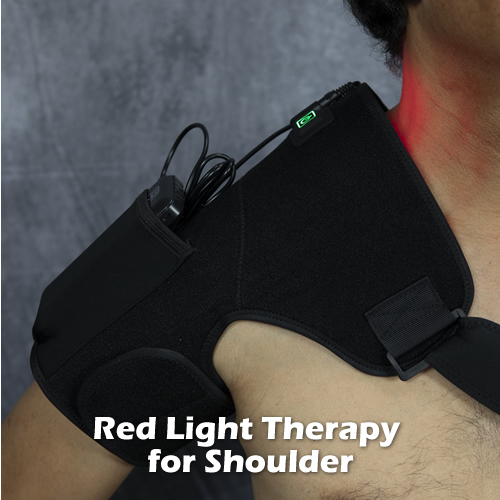
Lastly, I apply red light therapy.  I’m an advocate of this therapeutic technology and have written articles about it.  Red light therapy is actually an electromagnetic waveform (600-880 nanometer wavelengths) that appears red to the human eye.  It’s not the red you get from shining a light through a red lens; it’s a specific waveform in the electromagnetic spectrum generated from an LED (light-emitting diode).  The device I use uses three LEDs, one of which emits a waveform closer to infrared and therefore does not appear to be red as it is invisible.  The electromagnetic energy is at a frequency that gets absorbed by cell mitochondria and other structures, which can result in changed oxidative states that lead to cell signaling that initiates reparative processes, such as increased ATP production and increased membrane permeability.  This lessens inflammation and stimulates healing.

red light therapy device

I anticipate my trigger fingers to fully recover, to pre-injury status.  I will continue to do these therapies, as I feel they are partly responsible for my good results.

BOTTOM LINE

Prevention is the best cure:  if you know you are going to be using your hands a lot, such as starting on a do-it-yourself project involving power tools and hard gripping, know that this can cause trigger finger.  Do what you can to minimize the stress to your hands—take frequent breaks; don’t overdo it/ don’t hold a power grip for more than a few seconds; and rest and stretch your hands often.  Don’t rush it.  Trigger finger creeps up on you, and by the time you notice it, it is too late.  The presentations are different from person to person, depending on age, health, fitness and so on.  I am lucky as my condition is resolving; others are not so lucky and wind up getting surgery and permanent percent loss of hand function.  So make sure you keep prevention in mind.  If you do get it, try the treatment methods for trigger finger described here that have worked for me.

How Pulsed Electromagnetic Field Therapy (PEMF) Can Improve Your Health

How Pulsed Electromagnetic Field Therapy (PEMF) Can Improve Your Health

Pulsed electromagnetic field therapy, or PEMF is a safe, effective treatment for many types of health conditions.  Some people use it for arthritic pain, while others use it for fibromyalgia, insomnia, and even chronic lung and cardiovascular illnesses.  Unlike medicine and surgery, PEMF therapy doesn’t have dangerous or serious side effects. 

There are many types of PEMF machines on the market, direct to consumer so it doesn’t require multiple trips to the doctor for treatment.   The beauty of this is that PEMF can be used on-demand: whenever you have an acute flare-up or especially bad day; or, for daily maintenance of health.

And, since it is safe and doesn’t involve drugs, PEMF therapy can be used with other interventions that you may be currently using to reduce pain and inflammation, which leads to faster healing and recovery.

You may be wondering at this point, “how does Pulsed EMF work?”

PEMF – The Basics

As the name implies, PEMF therapy uses magnetic pulses that it sends into the body to speed up healing of weak cells, and therefore recovery.  The human body emanates electric charges (think brain and nerve impulses and charged molecules and ions like Ca+, Mg+, H+ and OH-) and just like inanimate things that require electricity or batteries to keep them going, your body needs “recharging” once in a while.  Several trillions of cells comprise your body, depending on how big you are, so it’s not surprising that many of them can break down at any time.

Healthy, normally-functioning cells have a balance between positive and negative charges (see image below).  As these charges flow inside and between adjacent cells, electrical currents are produced.  This electrical flow is the means by which nutrients and waste products enter and exit through cells, via special structures called ion channels.   For example, when nerves are stimulated, they “depolarize,” where the negative and positive charges rapidly exchange position (inside vs. outside the membrane) and move down the nerve.  This is called an “action potential” and is the actual means by which our nerves are able to contract our muscles, control our organs and send sensory signals to our brain.

cell membrane potential

When this critical flow of electrical currents through cells is disrupted, cells don’t carry out their functions as well as they need to.  This leads to illness and symptoms.  Things like trauma, disease processes, and exposure to toxins can alter these electrical signals.  Pulsed electromagnetic therapy uses the same, natural frequencies produced in the body to give these cells a boost in energy. This restores the positive and negative charges in the weakened cells to their ideal state so they can once again function normally.

You may have heard of the dangers of electromagnetic fields (EMF) or electromagnetic radiation, especially if you follow alternative health websites. The frequencies used for PEMF therapy is nowhere near that of harmful EMF such as X-rays, gamma rays and microwaves.  PEMF generates non-ionizing radiation, identical in frequencies already emitted by your body and therefore generally accepted as harmless to humans.

Health Benefits of PEMF therapy

There are many research studies that support the use of PEMF therapy for a wide range of disorders, from acute (recent; active) injury or health problems to chronic pain.

According to studies, PEMFs are useful in treating post-surgical pain; pain related to diabetic neuropathy, joint pain from osteoarthritis, and chronic lower back pain.  PEMF therapy has also been shown to increase the healing rate of nonunion fractures, and even increase bone density in those who have osteoporosis.  PEMFs ability to increase microcirculation to tissues is likely a factor.

Perhaps most impressively, PEMFs can help your body decrease inflammation, which is present in many chronic conditions including fibromyalgia, inflammatory arthritis, chronic fatigue syndrome, and autoimmune disorders such as Chron’s disease.

There are even studies that found PEMF therapy to have measureable, positive effects on those battling depression and anxiety, with long-term, daily treatment.

Conclusion:  Pulsed EMF is a safe alternative for healing

If you are battling disease and/or pain, consider trying Pulsed EMF.  It has scientific research going back over 40 years that support its many health benefits, and it is comparatively safe/ low risk to most individuals (those with heart conditions and implants should check with their doctor first).  PEMF machines produce electromagnetic fields at the same frequency as the human body’s, so cells respond to them in a positive way.

PEMF machines available for home use are low intensity, so there is no danger of excessive exposure that could lead to significant adverse side effects.  Treatment time can vary from just a few minutes to continuous, overnight use, depending on the problem being treated.  The machines offered on our site are established leaders in PEMF technology, from the Russian-manufactured Almagia line to the HTC products—FlexPulse, BioBalance and TeslaFit lines.

Bio Balance Pulsed EMF

There are some higher intensity machines (TeslaFit) meant for use in clinics that are more effective for long-standing, chronic conditions rooted deep inside the body; but most conditions can benefit from the lower intensity units.

The FlexPulse is a low intensity, portable PEMF unit available direct to consumer.  It was developed based on NASA stem cell research done over 15 years ago, and decades of research on brain entrainment (synchronizing biological frequencies) and neurofeedback.  By increasing stem cell (cells that can turn into any type of cell) production, the FlexPulse promotes safe, natural regeneration with no side effects.  Specific, pre-programmed frequencies mimic brain patterns to promote relaxation or alertness, enhance sleep, and relieve pain depending on your treatment goals.

The FlexPulse is a compact, wearable system that comes with two small pad applicators making it easy to use anytime, anywhere.   The system comes with a 30 -day satisfaction guarantee and a 2-year International warranty, making it a no-risk healing alternative.

Check out our full line of Pulsed EMF machines here.

Watch the video below where I explain how Pulsed EMF can re-energize weak cells and help restore health.

A Look into George Floyd’s Cause of Death, From an Anatomical Perspective

A Look into George Floyd’s Cause of Death, From an Anatomical Perspective

On May 25, 2020 an African-American man named George Floyd was apprehended by four police officers in Minneapolis, Minnesota.  During the apprehension, a police officer named Derek Chauvin placed the handcuffed Floyd prone on the street, then knelt over him, placing his left knee on top of Mr. Floyd’s neck.  From the image circulated in the media, it appears that Chauvin exerted his full weight over his bent knee.  He kept his knee in position for 8 minutes and 46 seconds despite please from Floyd that he could not breathe.  Tragically, Mr. Floyd died at the scene.  Two autopsies on Mr. Floyd ruled the death a homicide.  The county version “revealed no physical findings that support a diagnosis of traumatic asphyxia or strangulation” while a private autopsy ruled he died of “asphyxiation due to neck and back compression.”  Asphyxiation is the deprivation of oxgyen to the body, resulting in death.  Chauvin was fired and arrested shortly after the incident, and charged with 3rd degree murder and second degree manslaughter.

This tragic and awful case spurred protests and civil disobedience throughout major cities in the U.S., against systemic police brutality and racism towards African-American men, which continues as I write this.  It is also a case that has drawn interest in the medical and forensics community, as to how exactly George Floyd died.  This is important to investigate, as it has consequences for the trial and also provides information to police forces to help them determine which types of restraints should and should not be used.

Before discussing the medical aspects of Mr. Floyd’s death, I want to emphasize that the most important thing about this incident is that a man unnecessarily lost his life to a trusted law enforcement officer, in a most inhumane way.  It is especially bad because of the systemic racism element to it, and the fact that Chauvin had several opportunities to get off of him in time after being warned by another officer at the scene and multiple witnesses, but failed to do so.  This was a textbook lesson on how not to subdue a person, and the price of this lesson is going to play out for weeks to months and will be costly to society in more ways than one.

That being said, I will discuss the anatomical and physiological factors involved in Mr. Floyd’s death.  There is some controversy over how he died, which will determine what sentence, if any, Derek Chauvin will receive. 

As you will see, the neck contains several structures directly tied to sustaining life, which is why the neck is a logical target when it comes to martial arts/ self-defense, and yes, murder.

Mr. Floyd was lying prone (stomach down) with his hands handcuffed behind his back and neck turned to his right.  So when Chauvin placed his knee over his neck it contacted the lateral (side) aspect of his neck and some of the anterior portion.  The critical structures found at the side of the neck include the vertebral arteries, cervical nerve roots, carotid arteries, jugular veins and lymphatic vessels.  Attached to the carotid artery is the carotid body, a cluster of special cells that detect oxygen saturation levels in the blood leaving the heart, and passes the information to the brain, which the brain uses to regulate heart and breathing rates accordingly.

Inside the cervical spine (the neck bones) there is the spinal cord which is comprised of the neuron axon bundles that control body movement and sensation; and lying just outside and against the cervical spine is the sympathetic chain ganglia, which play a role in innervating the heart, lungs, adrenal glands (adrenaline secretion) and other organs during “fight or flight” moments of stress.

The critical structures found in the anterior neck are the esophagus, trachea (wind pipe), larynx and thyroid gland.  Also present but not critical to life are the vocal cords.

The contact area on Mr, Floyd’s neck, based on the typical size of a bent knee, was about 4 square inches.  Chauvin weighs about 170 pounds, so I estimate that the force placed on Mr. Floyd’s lateral-anterior neck was (.9)(170 lbs)/4 in sq. =153 lbs/ 4 in sq. = 38.25 lbs./in sq.  So imagine four, 40 pound dumbells stacked and resting on the side of your neck for nearly 9 minutes.

What I believed happened is blood flow to the brain was cut off, making Mr. Floyd unconscious after a few minutes, as would happen in a martial arts choke hold.  We can assume the loss of consciousness due to restricted blood flow to the brain occured the moment he stopped talking.  Chauvin kept his knee in place for nearly 3 minutes after Floyd stopped moving.  If there was any chance of resuscitation, that chance ended with this additional time of compression.

The pressure also activated Mr. Floyd’s sympathetic chain ganglion, which caused a surge of adrenalin– a neurotransmitter secreted by the adrenal glands of the kidneys that prepares the body’s response to stress.  This increased his heart rate, increased blood pressure by vasoconstricting his arteries; dilated his pupils, and attempted to increase breathing rate.  However, with Chauvin’s knee on Floyd’s neck, the increased respiratory rate did not result in more oxygen getting to the body and the diaphragm, the sheet of muscle that pulls down and expands the lungs for breathing may have spasmed as it fought against the air blockage.

Most of Chauvin’s knee was over the side of the neck, but the front (anterior) was subject to some compression.  This likely partially collapsed the trachea, restricting air flow.  Mr. Floyd was initially able to express his inabilty to breathe, suggesting that his trachea was still patent, but his words became less and less as the seconds ticked.  Perhaps Chauvin’s knee shifted forward with his weight, slowly closing off the trachea.  This makes sense, as the neck has a curved contour which would promote such slipping.

As the knee hold persisted cutting blood flow to his brain, Mr. Floyd’s blood rapidly turned acidic as all oxygen was used up (the brain has a high metabolic rate and therefore a high rate of oxygen consumption) and CO2 levels were rapidly rising (CO2 is a byproduct of cell respiration).  This may have affected his speech center, which is supported by the fact he stopped talking a few minutes after the knee hold.  The apneustic (breathing) center in his brainstem was starting to break down as well from the hypoxia (insufficient oxygen), hampering his breathing further.

It’s not clear at what point Mr. Floyd expired.  Coroners define time of death at the point when brain activity ceases.  But the nature of Mr. Floyd’s death was so prolonged, it is likely he experienced irreversible brain damage and would have been in a vegetative state even if Chauvin had gotten off of him a little earlier and Mr. Floyd was given immediate medical attention.

One of the tell-tale signs of asphyxia by strangulation is ruptured blood vessels in the conjunctiva (whites of eyes) and face, called petechial hemmorhaging.  This information, as far as I know, is not available to the public.  If there was no evidence of this, I can see why the county autopsy reached the conclusion that he did not die of asphyxia.  In this case, it would suggest that Mr. Floyd’s primary cause of death was something else; perhaps sudden stoppage of the heart due to a breakdown in the cardioregulatory system.  But, it is not clear if you can die from asphyxia from “gradual” strangulation and not have petechial hemmorhaging.  It could also be that death was from a combination of both asphyxia and heart stoppage.

The autopsy did not mention fractured cervical vertebrae, which I could see happening if it were a smaller-framed person.  The force Chauvin exerted on the neck appears enough to break one or several neck bones, especially if the person had osteopenia (bone thinning).  Fractured neck vertebrae often result in spinal cord damage and paralysis.

Conclusion

George Floyd’s tragic death is a wake-up call for police forces across the country.  Knees to the neck to restrain someone must be prohibited.  There are too many ways for this to go wrong, given the high concentration of structures critical to maintaining life that reside in the neck.  I understand that safety to police officers is important and fully support it, but it must not be accomplished by jeopardizing the safety of the person being restrained.

What Causes Low Back Pain?

What Causes Low Back Pain?

Low back pain continues to be a problem for many people. If not you, then most likely several people you know:  neighbors, co-workers, friends and relatives. That’s what the statistics tell us.

Since it is so prevalent throughout the world, “what causes low back pain?” is a question millions of people want answered. Is it normal and expected as we age? Is it genetic? Will you need to get surgery? As you can expect, the answer is different for different people. First of all, the pain is not identical from person to person. Some people get low back pain on the right side; some get it on the left side. Some get low back pain into the hip.

In this post, I will do my best to help you understand what causes low back pain and at the end of the article provide you a tool to pinpoint what is causing your particular low pain.

The Lumbar Spine and Its Significance

Your low back or lumbar spine engineering-wise is your body’s lynch-pin– along with your pelvis, it connects your upper body to your lower body and is tasked with balancing and moving your torso. If you injure your low back it can put you out of commission: any attempt at moving places a load on your low back and makes pain worse. In extreme cases it is even painful to take in a deep breath! Acute low back pain can instantly stop a 250 pound football player in his tracks; that’s the power it has.

While most cases of low back pain self-resolve over a few days, about 20% of them become chronic, or recurring. For some, it strikes every couple of months; for others, it’s enough to impede their daily activities and quality of life. In fact, low back pain is said to be the number one reason for lost work days (disability) in industrialized nations, and therefore lost productivity.

The reason why low back pain is so prevalent is a societal phenomenon, made possible by evolution. You see, humans are the only bi-pedal animal on the planet. Dancing bears and meerkats don’t count because although they can walk a few steps their pelvic design is still quadri-pedal (walking on four limbs). When nature selected homo sapiens to be bi-pedal, it freed up his arms to carry things–heavy things. Carrying and lifting things and bending the low back places a tremendous load on the lumbar spine, and like any machine, the components bearing the most stress will be the first to break down.

Secondly, the invention of the chair and desk. When our ancient ancestors roamed the plains and forests 30,000 years ago there weren’t any chairs around to sit in for hours, placing pressure on the low back and weakening the postural muscles of the spine. Today, many jobs require sitting at a desk in front of a computer, doing just that. Also, food these days is abundant and much less nutritious causing humans to gain excess weight, placing constant stress on the low back throughout the day.

Causes of Low Back Pain

The vast majority of low back pain cases are mechanical in nature; meaning caused by a breakdown of some physical component of the lumbar spine. These components are the muscles, fascia (muscle sheath), ligaments and tendons; vertebrae, vertebral joints, and discs (which are technically ligaments). If the dysfunction causes compression of nerve roots, then nerve pain is involved, which usually means shooting / radiating pain and/ or numbness down the buttock to lower extremity; sometimes as far down to the sole of the foot.

Here are the main categories of mechanical low back pain:

Congenital Malformations

Sometimes there are abnormalities in the development of the spinal column which interfere with proper movement and balance placing excess stress on soft tissues and sometimes nerves, generating pain or constant stiffness and aches, and loss of range of motion/flexibility. Examples include fused vertebrae (two adjacent vertebrae fused together instead of forming a joint); scoliosis; spina bifida, pars defect, hyperkyphosis (hunchback); and hyperlordosis (swayback).

Injuries

Low back pain is often due to injuries to tissues: sprains to ligaments; ruptured intervertebral discs from a herniated or prolapsed nucleus pulposus (jelly-like shock absorbing substance in all discs); strains (tears, small and large) to muscles and tendons; muscle spasms, and fractures. These can be traumatic from a specific incident such as a sports injury, or can be cumulative over time, often years, from performing a certain movement repeatedly or sitting/slouching causing gradual degenerative disc disease. With acute tissue injury, the inflammatory response is initiated, which is responsible for the pain generation.

Degenerative Changes

Joints wear out over time. Most doctors will tell patients their condition is from “normal” wear and tear. But that’s not accurate. For some people, joints deteriorate at an abnormally fast rate, mainly due to lifestyle factors under their control. I’ve seen X-rays of 60 year-olds look much better than 30 year-olds, on many occasions.

Factors that promote lumbar spine degeneration include:

  • Being overweight
  • Genetic predisposition
  • Sedentary lifestyle/ lack of physical activity
  • Sitting frequently (airline pilot, police officer, truckers, data entry)
  • Heavy labor job
  • Contact sports, especially football
  • Occupation or recreation that involve hard landings (basketball, gymnastics, parachuting, etc.)
  • Previous injuries/accidents such as falls and car accidents
  • Poor diet (your body needs proper nutrients to heal tissues)
  • Smoking (smoking reduces oxygen to cells and may trigger inflammation)

Doctors use three terms to describe spinal degeneration:

  • Spondylosis when referring to the vertebrae as a whole;
  • Degenerative joint disease or DJD if referring to the vertebral joints: facet joints and intervertebral joints; and
  • Degenerative disc disease or DDD if referring to the intervertebral discs

In all cases, the joint surfaces of the vertebrae lose their smooth borders and form jagged bone spurs called osteophytes. You can have a lot of osteophytes in your spine and not feel pain at all. In fact, if you are over age 40 you probably have them yourself. But if the osteophytes get big enough to narrow the openings where nerves pass through, called foramen, problems start. This narrowing of the foramen is called spinal stenosis and can occur with the intervertebral foramen (IVFs), the small holes formed by adjacent vertebrae which nerve roots pass through; and also the central canal where the spinal cord and cauda equina reside. This can lead to shooting pain down one or both legs; numbness and tingling in the legs, and leg muscle weakness, atrophy and loss of sensation.

Since osteophytes do not resorb (shrink) and only get bigger with time the only option is spinal decompression surgery which involves shaving off the osteophytes to make more room for the nerves.

And there is another problem:  when spinal discs degenerate, they lose height (this is part of the reason why most people get shorter as they age). A healthy L5-S1 disc can be in excess of 1 cm thick while a degenerated one can be just 2-3 mm thick. When this happens, the posterior and anterior longitudinal ligaments that run down the front and back of your vertebral column slacken, or buckle, at those levels. Like osteophytes, buckled ligaments can cause stenosis, compressing or irritating nerves and causing the same neurological symptoms.

When the degenerative changes involve deterioration of cartilage, you have bone on bone contact. The cartilage in your spine is located in the encapsulated facet joints, located behind the vertebral bodies. This triggers inflammation, which leads to osteoarthritis. It’s the same process as osteoarthritis of the knees and hips, occurring in the spine.

Like knee osteoarthritis, people with spinal OA will feel burning pain in their lumbar spine especially when standing, and stiffness which is worse upon waking in the morning. Osteoarthritis is a chronic, degenerative disease that is best managed by lifestyle modification (anti-inflammatory diet, exercises, stress reduction). Those with severe cases sometimes elect to use prescription anti-inflammatory medication.

Non-Mechanical Causes of Low Back Pain

Less than 1% of low back pain cases are due to other factors, most of which are “red flag” cases that require immediate medical attention. These include pelvic tumors, kidney stones, metastatic cancer (usually from prostate cancer), infection, and endometriosis. A brain tumor is capable of causing sciatica-like symptoms if it affects the sensory neurons that go to the leg.  While rare, if you have low back pain that does not improve with physical therapy or rest, it is a good idea to see your doctor and get some tests done to rule out these conditions. 

Summary and Main Take Aways

If you have low back pain, chances are very good it will go away with rest. Apply ice for the first 1 to 2 days. If severe, you can try taking over the counter anti-inflammatory medications (NSAIDs) such as Motrin or Ibuprofen to knock down some of the pain. And if you are lucky to have a cryotherapy center near you, you can do a couple of visits to shorten the healing time.

If the pain lasts more than 4 days, then it usually means something is perpetuating it:  perhaps you are not resting it properly; are re-aggravating it; or have some kind of contributing factor such as a rotated vertebra or disc protrusion. These issues can be dealt with by visiting a good physical therapist or chiropractor. You can even do some home exercises and home therapy for low back pain on your own and still get great results.

While most cases of low back pain go away, it doesn’t mean that it will stay away for the rest of your life: 20% of people who get low back pain will experience it again in the future; either from a new injury/event or a flare-up of a pre-existing injury.  And remember, some cases become chronic (pain decreases, but the improvement plateaus and remains, with some days worse than others).  Your prognosis will depend on what is causing your low back pain (see below for a self-diagnosis tool) and other factors related to your medical history and daily activities.

When dealing with low back pain, besides focusing on reducing the pain think of what things caused it and eliminate those causes as best you can. It may mean:

  • Getting a stand up desk (standing puts less stress on your low back than sitting)
  • Losing some weight
  • Eating a healthier diet
  • Stopping smoking
  • Exercising more often
  • Reducing emotional stress in your life
  • Improving the ergonomics of your work station
  • Learning how to lift properly
  • Getting your back adjusted by a chiropractor periodically to improve joint movement and joint health

And lastly, remember that low back pain should noticeably and progressively improve each day after initial onset. If you notice that pain does not get better with rest, express your concern to your doctor: demand an X-ray, MRI and/or a blood test. Red flag cases like cancer are often misdiagnosed by doctors (dismissed) as general back pain due to spasms because doctors know that the medical literature estimates red flag cases to be 1% or less of all low back pain cases. Remember to be persistent; it is your life you are dealing with.

Below is a low back pain algorithm I created to help you diagnose your particular low back pain so that you can take appropriate action. Use the top one if your low back pain onset was sudden; use the bottom one if the onset was gradual. This is only a guide; always check with you doctor who can examine you, to get an accurate diagnosis.

Self Rehab Videos for Pain In Production

Self Rehab Videos for Pain In Production

Dear Readers,

As we approach age 40, one of the subtle, inevitable changes we experience is shrinking muscles and weaker joints. Since males on average have a higher percentage of muscle mass, the changes are more noticeable: less strength in the gym; it’s harder walking up stairs and harder to run. Shrinking leg muscles and loss of speed and agility are what cause great athletes like Michael Jordan and Joe Montana to call it quits.

What should this mean to you?

It means that if you want those future plans you have in mind to become a reality, you need to take care of your body today because it is changing, which demands certain adjustments in your daily routine to compensate. I know people whose life changed dramatically after blowing out a disc in their back. While injuries like this eventually heal, they often result in a percent loss of function and chronic pain. Those who got surgery didn’t fare much better most of the time, as surgery disrupts nature’s design.

With that, allow me to be one of your resources to help you improve and maintain your musculoskeletal health. Even if you currently don’t have pain or limited mobility, I hope you stick around because: (1) prevention is the best cure for disease; (2) your situation can change any day; and (3) you might discover something from one of my videos or articles that you can share with a friend who is in pain, and possibly save him/her from unnecessary suffering.

Products. I’d like to be transparent about this, so let me explain. But first — did you notice the Pain and Injury Doctor logo?   A quick glance and it looks like the Red Cross symbol. Look closer, and the logo is actually four arrow pointing inwards. Each one represents a component of self-care for musculoskeletal health:

1 – diet and nutrition
2 – rehabilitative exercise
3 – manual therapy
4 – home modalities

(The four arrows form a small person with arms and legs extended (X) to symbolize maximum flexibility).

PainandInjuryDoctor.com falls under telemedicine; a broad term that encompasses aspects of health care delivered over the internet.  You’ve probably heard of sites like WebMD, ShareCare and Mayo Clinic.  These are more than websites; they are a form of telemedicine.   They enable individuals like you to research symptoms, drug names, diseases and medical terms used by your doctor and get fast answers.

With PID, you get guidance on how to self-manage common, non-emergency forms of musculoskeletal conditions such as low back pain, neck pain, tendonitis and shoulder stiffness.  Yes, there are tons of videos on YouTube on this; some good and some not-so-good.   My intent is to explain the cause of common musculoskeletal conditions in simple terms, and special strategies I developed to get relief, even permanent relief, that involve the four components mentioned above.

The Big Idea of PID is that yes, you can cure common presentations of musculoskeletal pain faster than waiting it out by doing home rehabilitation.  I’ll share with you videos of the techniques I prescribed to patients when I was in practice, and still personally use to get relief when needed.  Some of these techniques involve using equipment.  Yes, I do sell them on my site PulsedEMF.com.  It’s obviously up to you to purchase them, and if you decide to do so, where to buy them.  My job is to show and explain what I believe will bring the best, fastest results in relieving pain but the action is up to you.  Rest assured, the products on my site are the select few out of nearly a hundred that I have tested over my 20 years in practice that I’ve seen actually work on pain sufferers.  In fact, I have many of these products lying around my home that I use regularly to treat friends and family.  They all have scientific and/or clinical evidence to back them up.  Think of them as investments in your health and quality of life.

I truly believe this is just the tip of the telemedicine iceberg, and greater things are yet to come as technology advances.  I believe that virtual assistants like Alexa, in the near future, will be able to project a hologram demonstration of exercises to do for a bulging disc; or be able to accurately diagnose a condition by scanning your body and reading your vital signs, right in your living room.

In the meantime, I welcome you to keep visiting The Pain and Injury Doctor online.  I am in the process of producing self-rehab video guides for the 15 most common presentations of musculoskeltal pain I’ve come across.  The ones for Low Back Pain and Fibromyalgia and Insomnia are completed.  Next up is Neck Pain.  Please share on social media, as there is a good chance someone you know can benefit from these free rehab videos.  They are taking longer than anticipated to produce, partly because of the Covid pandemic, and partly because of my high quality standards.  But when they are complete, I hope for these videos to help millions of pain sufferers across the world; especially those who do not have access to a doctor.

Also in the meantime, I plan to release more articles and YouTube videos on managing pain.  Some of the topics coming up include what to do about foot weakness and pain; a great, inexpensive home recipe for joint health; and a factor that might be affecting your health that you probably don’t know about.

Till then, take care, and stay vigilant until the pandemic is declared over.

Dr. Perez

P.S. You can ask me a question any time on my FaceBook page, or you can use the form at the bottom of the PID home page.

Receive a FREE, 30-Day Plan to Boost Your Health and Eliminate Pain!

Receive a FREE, 30-Day Plan to Boost Your Health and Eliminate Pain!

As a subscriber, you'll also learn the special methods used by experts in human biomechanics to fix body aches and pain the RIGHT way, long term. 

We'll also send you a Free eBook, Concepts of Self-Healing as a way of saying thanks.

Please check your email in 5 minutes to access your Special Report. Make sure to whitelist "[email protected]" in your email client (Gmail, Yahoo, Outlook, etc.) so that you don't miss this valuable information. One way is to add this email to your email Contacts.