Aneurysmal Bone Cysts

 (Updated 3/9/2021
An aneurysmal bone cyst (ABC) is a painful condition in which an aggressive, cystic (fluid filled cavity) lesion develops in a bone, usually following a fracture or trauma, sufficient to alter the venous circulation of the bone.  It can also develop from pre-existing bone tumors.   It is typically found between the ages of 10-30, with the peak age of 16.  The most common site is the knee region (proximal tibia), followed by the arm bones, spine, and upper leg (femur).  ABCs have also been documented in small tubular bones of the foot, and skull.

The symptoms of an aneurysmal bone cyst depend on the affected area.   A common presentation is relatively fast onset of pain that rapidly increases in severity over 6-12 weeks.

The  skin surface temperature over the cyst may increase, a hard, bony swelling may be felt , and movement in an adjacent joint may be restricted.

Spinal lesions may cause radiating nerve pain or quadriplegia, and patients with skull lesions may have moderate to severe headaches.

Although aneurysmal bone cysts are relatively rare, I decided to address this disorder because the symptoms may be wrongly interpreted as a sprain or strain, especially for ABCs that affect the posterior elements of the spine.   A wrong diagnosis will lead to the wrong treatment prescription, such as ongoing chiropractic adjustments or physical therapy, which can worsen the condition.

If you have a teenager who complains of sudden, focal pain around a bone, frequently the knee, it’s best to take your child to his/her primary care physician, who should order a skeletal X-ray.  X-rays are the preferred imaging study to diagnose ABCs.

aneurysmal bone cyst knee xray

There are several treatment options for aneurysmal  bone cysts.  Surgical curettage, which is basically scraping out the cyst; embolization of the cyst (blocking its feeding veins and arteries), and injection of drugs to inactivate the cyst.

Aneurysmal bone cysts are not serious conditions at the same level of osteosarcoma, but they should be dealt with as soon as possible to prevent worsening.  They reduce the strength of bones and often lead to bone collapse/ fracture that could be dangerous.

There are surgical interventions that involve reducing the cyst.  You may want to try pulsed electromagnetic field therapy (PEMF) over the lesion.  PEMF is the external application of electromagnetic field energy into the body.  The field is low frequency and low amplitude and is similar to the natural magnetic fields produced by your body.  It is known to enhance cell membrane transport and improve red blood cell flow within the small capillaries.  For more information on Pulsed EMF, click Here.

How to Tell if Your Pain is from Arthritis

Arthrite rhumatoide Source: http://nihseniorhe...

Image via Wikipedia

If you have deep, achy pain in a joint that doesn’t go away with time it’s likely some form of arthritis.  Lower back pain can be arthritic in nature (as opposed to muscle) if it behaves similarly– deep, achy, doesn’t seem to ever go away entirely; and you have a history of repetitive, physical impact to your body such as construction workers/ laborers, martial art practitioners and gymnasts.  However, with rheumatoid arthritis (explained below) no history of trauma is necessary.

Most people associate arthritis with the elderly.  But did you know it can affect younger people as well?

The word arthritis translates to “joint inflammation.”  There are several forms of arthritis, the two most common types being rheumatoid arthritis and osteoarthritis.

Diagram of a synovial (diarthrosis) joint.

Image via Wikipedia

Rheumatoid arthritis (RA) is an auto-immune disorder, where the body’s white blood cells attack the synovium of synovial joints.  Synovium is the inner lining of a synovial joint— encased (capsular) joints like in the spine, hips, knees and shoulders.  The synovium produces synovial fluid, which lubricates the inner surfaces of the joint as it moves.

People suffering from rheumatoid arthritis will have  bouts of severe joint swelling, redness, heat and pain.  RA is typically bilateral; meaning, for the extremities it strikes both sides, not just one.  It is an awful condition that can can severely impact mobility.  Those with rheumatoid arthritis in the knuckles of the hand will have difficulty gripping things and doing fine dexterity movements such as writing and buttoning a shirt.  Areas commonly affected by rheumatoid arthritis are the hands, spine, and feet.

Currently, drugs are the only medical treatment for RA including non-steroidal anti-inflammatories– NSAIDS (aspirin, Motrin, Tylenol, Ibuprofen), corticosteroids and immunosuppressives.

Palliative care to manage pain levels include acupuncture, yoga, and chiropractic.  Cannabis (medical marijuana) is a popular choice for many sufferers.

Osteoarthritis (OA) is described as joint pain and inflammation from wear and tear of the cartilage.  It is usually found in people who have suffered repeated joint trauma, like football and basketball players.  Marathon runners can develop it in the knees and hips as well.  OA is found in the weight bearing joints of the feet, ankle, knees, hips, lower spine and neck.

When repeated trauma impacts a weight bearing joint, over time it creates tiny fissures in the cartilage, which thins the cartilage and exposes the bone underneath.  From there, the bone forms bone spurs called osteophytes.  Radiologists describe these changes (disc degeneration plus osteophytes) as spondylosis of the joint.

Both types of joints can be painful, with RA being more acutely painful.  The symptoms are pain, joint stiffness and reduced range of motion.

MAKING BONE BROTH SOUP FOR ARTHRITIS PAIN:

In both cases, eating a low-inflammation diet will be helpful:  reduce sugar intake including high fructose corn syrup; limit carbohydrates to 150 grams per day; emphasize protein and fat, especially fish with high Omega 3 content; nut oils (walnut, almond, Brazil nuts), virgin olive oil, and raw plants (green leafy salads, lightly steamed vegetables).   Raw milk and butter from grass fed cows, and coconut oil are also good for an anti-inflammatory diet.

Eating  bone broth soup every day will give your body the constituents for rebuilding cartilage:  Visit your local butcher and ask for the large beef and pork joints.  They can saw these in little pieces in the back and give them to you in a bag; they price them pretty cheap; too!

You’d be amazed as to how cow/ pig knee and hip joints look very much like a human’s.  If you want to understand the anatomy of these joints, this is a great way to do it.  You will see and feel the smoothness of the cartilage on the femur head.  You’ll see the anterior and posterior cruciate ligaments (ACLs and PCLs).  You will see the patella, fat pad; and collateral ligaments, condyles and meniscus.  Make sure to eat all of these parts in addition to drinking the broth to get all the proteins needed to rebuild tendons, ligaments and cartilage.

MAKING THE BROTH:

Add a couple of pieces of joints into a tall pot; fill half-way with water; add salt.  For extra bone-building strength add some eggshells (from cracked RAW eggs– You want the inner shell membrane to be intact; with boiled eggs the nutrient-rich membrane sticks to the egg).  Bring to a strong boil; reduce heat to low and cook for 60 minutes.  Steep out the eggshells, and drink the soup.  Eat any pieces of tendon stuck to the bones, and eat the bone marrow as well.  You can add spinach to the soup the last minute of boiling for more variety. (chicken and turkey carcasses and whole fish bones work well, too).

The calcium and cartilage nutrients you get from this bone broth can help your joints feel better.

HOME THERAPY FOR ARTHRITIS

Arthritis pain comes from inflammation and stimulation of pain receptors in damaged cartilage by the inflammation and also by physical abrasion.

Pulsed electromagnetic field therapy (Pulsed EMF or PEMF) can help by energizing the affected cells.  The electromagnetic field produced by PEMF machines are similar to the naturally-occurring fields produced by the biological activity of your cells; adding an external source helps those cells function better.  The few cells that exist in the cartilage and ligaments and tendons have more energy to synthesize matrix and collagen which goes towards joint rebuilding.  Use in conjunction with red light therapy for maximum effect.  Red light at around 660 nm wavelength initiates photobiomodulation, which also gives cells more energy to function, much like photosynthesis in plants.

For temporary relief, heat works best with chronic pain and stiffness.  An infrared heat lamp is a good choice, as it is easy to set up and penetrates all the way down to the affected tissue, unlike a hot pack which only heats the skin.  Heating is not advised for rheumatoid arthritis when it is in a flare-up, hot and acute phase.

As far as medical care, joint replacement is a more drastic option but can effectively reduce pain while preserving some joint functionality.  There are also experimental procedures that involve culturing cartilage cells in a lab and injecting them into the joint space, hoping that they will bond to existing cartilage and thicken, but results are mixed at this point.

If you don’t have arthritis, do things that will help prevent you for getting it.  Build up your joint strength with the diet mentioned above, and avoid repetitious trauma to the weight bearing joints (activities that involve jumping and landing on a hard surface).  Strengthen surrounding ligaments of your knee and hip joints with exercises like weightless squats, knee bends, and simple uphill hiking.

Your joints are the most neglected part of your body; we often take them for granted.  But when one is injured or develops arthritis, you will realize very quickly how important they are to your happiness and well-being.  Don’t wait until it is too late; strengthening and nurturing your joints should be a definite part of your fitness routine.

When You Get Whiplash Neck Pain from a Car Accident

When You Get Whiplash Neck Pain from a Car Accident

Ever get rear-ended while waiting in traffic or at a stop sign?  It’s a scary moment:  the calmness of being out in your car is viciously interrupted by screeching tires and a loud crash and bending metal.  It’s over in two seconds, but a lot happens to your body in that one second.

If a high speed camera was available to record your mishap, it would show your body violently moving forward with the car, back sinking into your car seat, your head slowly arching backwards at an unnatural angle; your neck muscles tense up; your head coming to a  stop in its backwards path and then reversing directions to move forward again, through a complete arc until your neck is fully flexed forward in an unnatural position; coming to a stop again, and then reversing directions and fully extending a few degrees less than before, and then reversing and flexing forward again, then righting itself.  That is your basic whiplash injury.

You’ll notice your neck become stiff, but very gradually.  You may be a bit dazed, and have a headache come on in about an hour.  You’ll likely feel tired.

As the day wears on, your neck is getting increasingly stiff and painful.  You may feel the onset of soreness in your upper shoulders and upper back; even your lower back.  You may have pain in your chest wall where the seatbelt dug into.

2-3 days after the accident your neck will reach maximum stiffness.  You will have difficulty turning your neck.  You’ll find yourself turning your whole body in order to see to one side.

So what is happening?  You have sustained what’s called a cervical acceleration-deceleration sprain strain injury; commonly known as whiplash.  Car accidents are a common cause of whiplash, but they can occur on roller coasters and similar jerky rides, horse riding, sky diving, and even wild dancing.  When this happens, tiny tears develop in the muscle tissue and fascia (muscle covering) which starts to release the inflammatory products of swelling.  The swelling is gradual, like a pinhole leak, which explains why it takes 2-3 days to reach max pain.  What makes a whiplash worse than other sprain strain injuries is that, due to the flexibility of the neck, spinal ligaments also incur damage.

When your neck flexed violently forward, the capsular ligaments and interspinous ligaments likely got damaged.  The capsular ligaments hold your neck bones together from the back; the interspinous ligaments hold them together at the spinous processes (the bumps you feel along your spine are the tips of the spinous processes).

interspinous ligament rupture

When these ligaments injure, the swelling goes inside the joint space, building up pressure essentially splinting (immobilizing) the joint.  In severe cases, the ligaments can rupture (tear) causing dangerous instability, and you have a very serious condition that requires a visit to the ER.  A head halo support or neck brace is usually attached to prevent the instability from causing damage to your spinal cord.

TREATMENT:

Obviously, it’s a good idea to go to the hospital if you were involved in a significant car accident and feel you’ve been injured.  The ER doctor will rule out serious conditions like ligament rupture, bleeding in the brain (subdural hematoma), and bone fractures.  Once those are ruled out, he/she will diagnose you as having a sprain strain injury and will usually prescribe pain meds (anti inflammatories and muscle relaxants).  You will be given home care instructions.

With whiplash, the goal is to first reduce the pain and swelling.  You will do this by applying an ice pack to your neck.  Here’s a YouTube video on Whiplash Home Care – icing and stretches that illustrates the information to follow.

Buy two gel ice packs at your local drugstore (9″ x 6″ size); put in freezer.   Make a cervical roll using a small bath towel or hand towel:  roll it up tightly into a cylinder 1′ long with a radius of five inches.  Place it on the floor.

Place one ice pack on top of the roll, and one right under (next) to it.   Put a kitchen towlette on top of the ice to prevent iceburn.  Lie down, face up with the center of the back of your neck on top of the cervical roll/ice pack.  The other ice pack is for your upper back muscles.   Put a pillow under your knees for comfort, dim the lights and rest for 20 minutes.  Repeat this every two hours, for 2-3 days.

On the second day, while icing slowly turn your neck to the right as far as you can, then to the left, then to center.  Then, arch your neck and touch the carpet with the crown of your head and hold for 20 seconds.  Repeat these motions ten times.  Do this for each of your icing session, still at 20 minutes every 2 hours.  This helps to regain full neck range of motion.

On the 4th day, alternate ice with 10 minutes of moist heat using a hot water bottle with 150 degree water heated on your stove, and a wet face towel for heat conduction, on your neck and back muscles.

As the pain decreases, engage in active stretching exercises.  About two weeks post crash, or when the pain has gone down 90% and you have full range of motion in your neck, do neck strengthening exercises.  It is important to do these rehab exercises as they help align the reparative tissue in the axis of contraction of the muscles.  This will help reduce the chances of chronic pain and loss of range of motion following the accident.

Since the original publication of this article, I’ve researched a new modality called Pulsed Electromagnetic Field therapy, or Pulsed EMF, or PEMF for short.  The link explains it in detail, but basically  it is the external application of low frequency, low amplitude electromagnetic fields, similar to the natural EM fields your body produces, to impart energy to them.  EM fields are used to drive movement of molecules in and out of the cells, which includes nutrients, waste products, proteins and other factors involved in life processes.  When cells are sick or injured, this process is not efficient.  Pulsed EMF imparts a boost of energy (in fact, it is considered “Energy medicine”) which helps injured/sick cells perform their biological functions more efficiently, promoting accelerated healing and improved symptoms (less pain).

Apply PEMF to your neck three times a day for 15 minutes, for 2-3 days following the accident.  You should experience a noticeable reduction in pain each time, an hour or so after treatment.

Wrist Pain and Treatment

Wrist Pain and Treatment

Wrist pain from overuse affects over 50% of people whose job requires frequent typing on a computer keyboard.  However, any job or activity that requires repetitious finger movement is capable of causing this type of wrist pain.

wrist tendon anatomy

The leading culprit is tedonitis, or tendinitis — inflammation of the wrist tendons; more precisely the flexor digitorum tendons which pass through the wrist.  As you move your fingers alternating between flexion and extension, which is essentially what typing is, the wrist tendons rub against one another.  The tendons are protected by a slippery sheath to allow smooth gliding during movement, but if this sheath loses its protective qualities, it can cause the tendons to inflame and swell.  Certain drug side effects and illnesses can cause this; check with your doctor.

The problem with tendonitis is that, by nature it affects tendons of muscles that are needed for work or play; for example the wrist for typing, the elbow for golf and tennis, and the achilles tendon for running.   The individual therefore continues in the offending activity until she can no longer tolerate the pain, and by that time a lot of microtrauma has occurred.  The microtrauma (tiny tears and fissures in the tendon and sheath) releases inflammatory products and attracts scar tissue formation, which makes them stick together even more.  The tendons undergo trophic changes (changes on the cellular level) and don’t function as well as before leading to chronic pain.

Prevention is the key for all cumulative trauma/ repetitive strain injuries– these painful conditions are totally preventable and don’t need to happen in the first place.  You don’t want to ignore prevention because tendonitis takes a long time to resolve once it is entrenched.  In some cases, tendonitis can lead to carpal tunnel syndrome, where the median nerve in the wrist gets squeezed and injured leading to numbness and weakness of the hand.

For wrist tendonitis from typing, make sure your workstation is ergonomically set up:

  • Use an adjustable keyboard tray to lower and angle down your keyboard at a level where you don’t need to contract your shoulder muscles to raise your arms above the keyboard.
  • Keep your upper arms in the same plane as your torso, and close to your sides (don’t type chicken winged).
  • Keep your elbows angled at about 95-100 degrees, wrists straight in line with the forearm or slightly bent downwards
  • Use a good chair with lumbar support and if necessary a foot stool to rest your feet on
  • Head up, ears directly over shoulders, eyes in line with center top 3rd of  computer monitor; shoulders relaxed.

Take 30 second to a minute mini-breaks if you are going to be typing more than an hour straight.

Stretch wrists periodically in flexion and extension; hold for 10 seconds; do wrist circles ten times in both directions.

Do shoulder circles ten times in both directions.

Do chest and neck stretches every other hour during the day.

Stand up and do wall angel exercises (50) at least twice during the day to counteract the tendency for neck and shoulder flexion during sitting.

TREATMENT:

1. Do ice massage every two hours during the day if your pain is acute (feels hot, swollen and in some cases, reddish skin).  Here’s a YouTube video of how to do it.

2.  Apply Red Light Therapy to your wrist.  The Red Light torch is great for this.  Press it gently but firmly into your wrist and depress the button.  Do three times/ day, 10 minutes each.  Research shows that red light therapy reduces inflammation, almost as good as a non-steroidal anti-inflammatory medications.

3.  To further accelerate healing, apply Pulsed EMF to the painful areas.

SUPPORTING EFFORTS

Eat an anti-inflammation diet for 2-3 weeks:  go heavy on fish oils; reduce grain intake; eat plenty of alkaline foods (green leafy plants).

Don’t do stretches when your tendons still hurt; introduce light flexion and extension stretches when the pain drops by at least 75%.

Do your best to reduce the activity that precipitated the tendonitis for 2-3 weeks.

When you have cured yourself of the pain, implement the preventive strategies mentioned above so it never happens again.

Sciatica – Ever Had a Pain in the Butt?

Sciatica – Ever Had a Pain in the Butt?

(Updated 3/29/2021)

No, I don’t mean a really bad boss or acquaintance.  I mean literally.

Buttock pain, medically called gluteal pain is a deep pain usually affecting one buttock.  The sensation is often described as an electrical-like shock shooting from the bottom area of the buttock down to the back of the same side leg.  Numbness and tingling is also often present.  When not shooting, it feels like a bruise and is quite bothersome.  Sciatica can make prolonged standing difficult, and even light walking.  Sitting is very uncomfortable.  Walking upstairs can make it hurt even more.  Slouching over tends to ameliorate the pain.

The pain is usually described to be in the cheek part (lower) of the buttock a couple of inches to the side and downwards, from the sacrum– the triangular bone at the base of your lower back that is connected to your pelvic bones on either side.  In some people, the pain continues down the back of the thigh to the calf, and sometimes the bottom of the foot.

In many cases, the patient cannot explain what caused the pain.  This type of problem usually is reported to have a gradual onset, getting progressively worse.

Sciatica, or pain down the sciatic nerve is usually the diagnosis.  The sciatic nerve is the largest nerve in the body.  It  forms from nerve roots that branch from the sides of the lower lumbar spine and sacrum.

However, the term “sciatica” describes a symptom and does not provide much clinical information.  Sciatica can be caused by a herniated disc that presses on a nerve root that partially forms the sciatic nerve; spinal stenosis, which is narrowing of the lumbar spinal canal to the point where the cauda equina, the long nerve roots that form at the terminus of the spinal cord, experiences irritation/compression; it can be caused by a pelvic tumor that encroaches upon the sciatic nerve (or in very rare cases, the part of the brain that controls the affected leg), and it can be caused by a condition called piriformis syndrome.

piriformis syndrome compression

Piriformis – Muscles of the Lower Extremity Anatomy Visual Atlas, page 8 (Photo credit: robswatski)[/caption]

The piriformis muscle is one of the small muscles that connect your hip bone (femur head) to the side of the sacrum and is used to externally rotate the leg (turn outwards).  The sciatic nerve passes between the piriformis muscle and adjacent muscles before it descends down the leg.  If the piriformis muscle is in spasm, it can squeeze the sciatic nerve causing deep gluteal pain; the piriformis muscle itself can generate pain if it is inflamed.

Piriformis muscle spasm can be caused by a hard fall on your seat that puts the muscle in spasm.  It can also develop when the pelvis is uneven; i.e. one hip is higher than the other when standing.  This can cause uneven pull on the gluteal muscles, causing the hip rotators to spasm.

SELF-TREATMENT FOR SCIATICA

If the pain persists for over a month or is unrelenting, see your doctor so that a herniated disc, spinal stenosis and tumor can be ruled out (you may get an MRI).  Once that is ruled out, you can do a couple of things to make the pain go away:

1.  Check the level of your iliac crests while standing in front of a mirror.  Place your fingertips flat on top of your hip bones; see if one is higher than the other.  Sometimes if the unevenness is large, you can see the high side and low side simply by looking in the mirror, and focusing on your belt or skirt/pant top — it will not be parallel to the ground, one side will be higher.  If this is the case, try inserting a heel lift in the shoe on the  side of the “lower” pelvic bone.  This will help even out the pelvis and remove strain to the gluteal muscles.  You can purchase an “over the counter” heel lift at most drugstores like Walgreens or CVS.  Check out this YouTube video that explains it:

Optionally, you can buy a pair of gel inserts for shoes, and use only one in the shoe on the side of the low iliac crest.  Make sure to wear the lifts for at least two weeks to see if they help.  Decreased pain or frequency of pain indicates that they are working.

2.  Slowly and gently stretch the painful piriformis muscle by lying on your back, raising the leg on the affected side and bending the knee.  Grab with both hands behind the knee and pull gently at a 45 degree angle relative to your body; you should feel a stretch in your gluteal muscle.  Hold the position for 30 seconds; repeat three times.  Watch this Sciatica YouTube video for a demonstration:

3.  Apply infrared heat with an IR heat lamp and red light therapy.  IR heat penetrates deeper, which is necessary to reach the nerve which is several inches deep, beneath layers of gluteal fat and muscle.

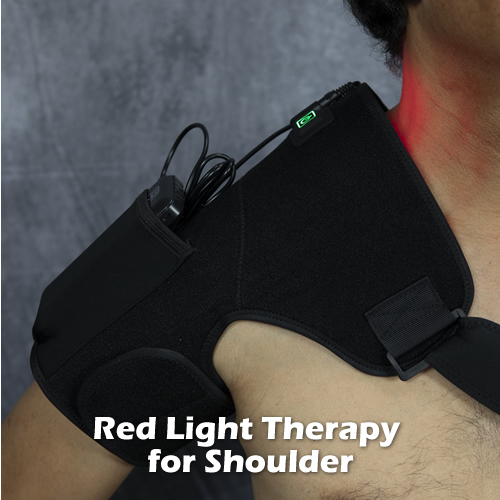
Follow up with a Red Light therapy wrap.  Red light invigorates cells and gives them a boost of energy.

red light therapy LED wrap

In summary, sciatica can be caused by several things.  Your prognosis is good if it is due to a bulging disc or piriformis syndrome, which can be managed by exercises, stretches and the therapeutic modalities I listed here.

If your sciatica is due to degenerative disc disease, spondylosis (bone spurring) and subsequent central spinal canal stenosis, unfortunately you are likely to have chronic, intermittent sciatica.  Some people with this presentation have found success with spinal decompression surgery involving removal of the posterior structures of one or several vertebrae (laminectomy), but it is a very risky procedure and can often cause new, low back pain problems.

Delayed Back Strain

Delayed Back Strain

Updated 3/29/2021

If you ever went to bed feeling fine but woke up with muscular back pain, it could be a number of things.

1.  Your mattress :  Cheap spring mattresses deform at the level of the body’s center of gravity when lying down, which is at the pelvis (buttocks/hip level).  Remove your sheets and take a look at your mattress:  is there a slight depression in the center?  If so, when you lie on your side, your pelvis sinks and forces your lumbar spine to arc downwards, compressing the joints and discs on the opposite side.

If you sleep on your stomach it puts your lumbar spine into hyper-extension (arcs sharply) forcing the facet (rear) joints of your lumbar spine together.  It also forces you to twist your neck, which rotates all the vertebrae all the way down your spine.

Sleeping on your back is the best position because it allows your spine to be in the most neutral position.  But if your mattress is old and has that pit in the middle, that can be enough to put a prolonged stretch on your back muscles as your pelvis sinks downward.  This can make your back feel sore in the morning.

Solution:  Get a new mattress.  I recommend the TempurPedic viscoelastic mattresses, which resist deformation (always spring back) and last a long time.  Basically, the mattress material pushes proportionally to the weight placed on it (more under the pelvis and upper back; less on the waist and lower legs) and rebounds so that it can do this every time.

If TempurPedic isn’t your thing (some users report feeling too hot in the summer on these mattresses), a high-quality spring mattress should work.

2.  You may have strained your fascia while exercising or engaging in other intense physical activity that required heavy use of the back muscles, such as rock climbing.  The fascia is the sheath that encases a muscle, protecting it as it lengthens and shortens inside the body, rubbing against adjacent muscles.  It may take longer for fascial strains to be noticeable as they don’t release as much inflammation as a strained muscle.   So, if you wake up and you have a very focal, sore spot on one of your back muscles that gets progressively worse, chances are you have a myofascial strain.  You probably incurred it the previous day doing a back exercise or other type of heavy exertion.

Solution:  Like all types of strains, fascia strains eventually resolve on their own, but it could be a couple of days and sometimes weeks of discomfort and problems with back mobility.  If you wish to eliminate your back pain sooner than later, consider getting a Pulsed EMF coil.

Pulsed EMF, PEMF for short, is the external application of a body-compatible electromagnetic field that energizes injured/ sick cells and one of the few medical devices cleared for home use that I highly recommend.  The magnetic field helps with molecular transport in and out of cells, which enables them to carry out their biological functions more efficiently; including healing, repair and protein synthesis.

Pulsed EMF has a number of reported health benefits such as pain reduction, faster fracture/injury healing, and better sleep.  Whole body PEMF devices are not cheap, but are a sound investment in your health.

More information on how Pulsed EMF can help you >>

3.  Your potassium levels are low.  Hypokalemia, or low potassium can occur if your diet is deficient in potassium, and/or you engaged in strenuous activity such as hard labor, a long hike or a long run and sweated quite a bit.  It is known to result in severe muscle spasms, sometimes involving one, particular muscle.  Potassium in ionic form (K+) is involved in nerve transmission and muscle contraction, so if you are low, it can cause dysfunction that manifests as a muscle contraction abnormality.

Solution:  Take Potassium supplements, about 500 mg/ day for several days until your muscle pain is resolved.

Bottom line, you don’t have to noticeably injure yourself to feel muscle pain.  Muscles are complex organs that can get damaged just like any other organ in your body if they are stressed in a way that interferes with their proper movement.  Potassium and other ionic imbalances, specifically calcium and sodium, can cause abnormal muscle function, and are therefore another possible cause of muscle pain.

How to Treat a Sprained Ankle

How to Treat a Sprained Ankle

Updated 3/20/2021

Most people have sprained their ankle at least once in their lifetime, me included.  It can hurt like the dickens and for some unfortunate people it can require a visit to an orthopedic surgeon and installation of surgical screws.

Thankfully, the vast majority of sprained ankles can be treated at home, and resolved 100% in time.

Most sprained ankles are inversion sprainsInversion refers to movement of the foot inwards relative to the ankle; i.e. “rolling inwards” of the foot while contacting the ground while bearing body weight.  Ankle sprains can occur when the person steps on or in something that s/he wasn’t expecting; for example a stone or a hole in the ground.  They also occur from sports activities.  Wearing the wrong kind of athletic shoes increases the chances of spraining an ankle; for example wearing running shoes while playing on a basketball court.

sprained ankle ligaments

Basic Ankle Anatomy

The ankle is a synovial joint (encased in a ligamentous capsule and lined with a thin layer of tissue called synovium, which produces synovial fluid for lubrication) that connects the lower leg with the foot (as a side note, synovial joints are the type involved in rheumatoid arthritis).  The articulating bones of the ankle are the tibia and fibula bones of the leg, and the talus bone of the foot.  Directly underneath the talus is the calcaneus, or heel bone which serves as an attachment point for some of your ankle ligaments.

All of these bones are all lined with cartilage, which absorbs shock and reduces friction in the ankle joint.   A major weight-bearing joint, the ankle evenly transmits the weight of the body through the bones of the foot.

There are several, small ligaments that connect the foot to the distal, lateral fibula, also called the lateral malleolus, or outer ankle.  Likewise, there are small ligaments that connect the foot to the distal, medial tibia, called the medial malleolus or inner ankle.  If the foot starts to roll in quickly as in an impending inversion sprain, mechanoreceptors (movement sensing neurons that send positional information to your brain) around the ankle sense the sudden movement and reflexively correct your ankle to avoid tissue injury.  Think of those times you almost tripped, but caught yourself; those are your mechanoreceptors in action.  If successful, you avoid spraining your ankle (it also helps to have good balance).

But if the mechanics and circumstances of the movement are such that they overwhelm this corrective reflex, the ankle joint will move past its maximum range of motion, which exceeds the ligaments’ load capacity resulting in a sprain injury.  Remember, ligaments connect bones to bones while tendons connect muscle to bone, so a sprain is injury of ligaments while a strain is injury of tendons.  They often occur together during injuries and are referred to “sprain-strain injuries.”

The primary function of ligaments is to hold bones tightly together while allowing some movement.  They do not have the specialized, contractile cells of muscles, but they can stretch (have elasticity) to some degree, due to the arrangement of the triple-helix collagen fibers within the matrix that act like a strong spring.

Below is a microscopic view of a human ligament (stained), showing tightly packed, spring-like collagen fibers.  The dark objects are fibroblast cells.

microscopic view of a ligament showing collagen fiber arrangement

In a sprain, ligaments tear to some degree.  Doctors describe the degree of ligament tears in grades, depending on severity:

Grade 1 – MILD.  Painful, minimal tearing of ligament fibers.  Ligament typically fully heals.

Grade 2 – MODERATE.  More painful, significant tearing of ligament fibers.  Can result in joint instability.

Grade 3 – SEVERE.  Pain, but sometimes not as painful as Grade 1 or 2.  Complete rupture of the ligament fibers.  If it’s a major ligament, it will require surgical re-attachment.

This sets in motion the body’s inflammatory response, which is a normal part of tissue healing:  a cascade of biochemical substances are released and the ankle starts to swell.  Maximum swelling is typically reached in about 2-3 days.  There may be a purplish color as well that gets progressively darker, which indicates leaking of small blood vessels pooling in the extracellular spaces.  Over time, the body clears the pooled blood out, and the color fades.

a sprained ankle showing internal blood pooling

Sprained ankle showing blood pooling in multiple areas.

Here’s the thing about inflammation:  it is necessary for proper healing, but it can overcorrect causing unnecessary pain and prolonged resolution of the injury.  This is why doctors advise using an ice pack during the intial stages to control the inflammation as cold constricts blood vessels making them less leaky.  Cold also slows down nerve pain signals to the brain.

Self Treating a Sprained Ankle

The first order of business is to do a simple assessment of your injury.  This will determine if  you need to see a doctor.  Note how much your ankle bent:  was it a short and quick sprain that your reflexes were able to correct before serious damage occurred?  Or, was your sprain such that you felt your foot bend completely inwards and had little control over correcting; and perhaps you felt and/or heard a snap?  When you grab your foot and attempt to move it, does it feel like there is no resistance, as though you ruptured all the ligaments, and it isn’t as painful as you would expect?

The second scenario is a Grade 3 sprain and warrants a visit to the doctor; the sooner the better.  These are the signs of ligament and possible tendon rupture, and even ankle or foot bone fracture.  With this kind of sprain there is increased probability of internal major vessel rupture, too, which can lead to a dangerous blood clot.

The first scenario is a Grade 1 or 2 sprain.  While the second scenario warrants a visit to the doctor, the following treatment can be done in both scenarios.

Old school treatment for soft tissue injuries is to use ice for the first 2 days, then heat.  But this is 2021, and there are new things available to treat injuries more effectively, which you’ll learn below:

1.  Get home, take off your shoe and sock if you are wearing them.  You should notice some swelling/ puffiness around your ankle.  Put two cups of ice cubes in a Ziplock freezer bag, and place over your ankle.  In the meantime, prepare an ice massager by filling a plastic cup with water and sticking a spoon in the middle, holding it in place with masking tape.   Put it in your freezer.   When frozen, pull it out of the cup by the spoon handle, and ice your ankle with it.

Don’t have the time for this?  Then check out this cheap ice massager on Amazon.  It’s even better.

The advantage of using an ice massager like this is that you can also use it to compress the swelling at the same time by pressing it into the tissue as you ice, firmly but not too hard.  This helps drive out the swelling.  Remember, R-I-C-E soft tissue injuries:  Rest, Ice, Compress, Elevate.  You can also use a plastic ice massager, like the one I’m demonstrating in this video which you can get on Amazon.  Do it for 20 minutes every two hours, for the first two days.

2.  Apply Red Light Therapy later in the day, and 3x/day for 10 minutes until it is fully healed.

Red light in the 660nm wavelength range is known to have healing effects via photobiomodulation.  Apply it around the lateral malleolus, pressing it firmly but not too hard into the tissue where the swelling is occurring.  You should feel the pain go down as you are doing it; it’s that fast.

red light therapy handheld device

3.  (OPTIONAL) If you want/need to make the pain go away faster and cut healing time in half, apply Pulsed Electromagnetic Field Therapy, or Pulsed EMF/ PEMF to your injured ankle daily until it is healed; 15 minutes, 2-3 times a day.  This is an option for you if your job demands frequent walking.  A Grade 2 ankle sprain can make walking pretty difficult for about an entire week.

Pulsed EMF is ideal for soft tissue injuries because it energizes cells that need more energy, i.e. cells in injured tissues.  PEMF devices emit a low frequency (safe) electromagnetic field that mimics the body’s own naturally occurring fields, augmenting them.  Electromagnetic fields  drive molecular movement in your body, which is also referred to as cell membrane potential (like voltage in a battery).  This boost of energy helps cells carry out their biological functions more efficiently such as nutrient transfer, protein synthesis, and waste removal.  It’s like speeding up a video so that it finishes faster, but its the healing you are speeding up!

You can use a Pulsed EMF coil belt device and wrap it around your ankle; or this hand- held Pulsed EMF device, which you can hold over the ankle or use an elastic band to hold it in place for 15 minutes.  They aren’t that cheap, but you’ll own equipment to heal injuries or pain you might sustain in the future, so they make a good investment in your health and well- being.

fibroblast cell secreting collagen

Animation of a fibroblast synthesizing and laying down collagen fibers.

4.  Move your ankle.  Passively (use your hands) and actively (use your leg/foot muscles) move your ankle starting a few hours after the injury, even if it is a little painful.  As your tissues heal, tiny cells called fibroblasts lay down scar tissue and collagen.  Moving your ankle through all its ranges of motion stresses the ligaments in the orientation of their fibers, which helps properly align the newly-formed fibers.   This is why immobilization (bracing and casting) is not recommended for these types of injuries.  However, you can wrap kinesiotape around your ankle with slight pressure to help reduce swelling.  Kinesiotape offers support, but also allows movement.

using a wobble board

A wobble board is useful for rehabilitating a sprained ankle.

As your ankle pain dissipates, consider using a wobble board to further strengthen/ rehabilitate all the muscles, ligaments and tendons that control ankle movement.

Bottom line, most sprained ankles can be effectively treated at home using the above approach.  It is important to ensure proper healing of any joint sprain-strain injury, because you risk re-injuring the same area more easily if the soft tissues heal sub-optimally, such as excessive scar tissue build up and loose ligaments that allow excessive joint movement, which can accelerate arthritis in the joint.


Website Launch

After months of planning, my new video blog PainandInjuryDoctor.com is finally up and running!

This has been something I’ve wanted to do since 2004 but my “real job” of being a busy doctor of chiropractic demanded most of my time; not to mention the addition of my son Sean who came into this world in late 2007 just when I was conceptualizing the format of this site.

However, things happen for a reason.  The delay in the launch was a blessing in disguise as I accumulated a vast amount of knowledge in musculoskeletal pain treatment and management over the past several years.  After being in practice for a while, eventually you feel like you’ve treated “everything under the sun.”  I believe I am close to this, as it’s been a long time since I’ve had a patient who presented with something I haven’t seen before.

This site will basically serve as a visual repository of what I know about treating common forms of musculoskeletal pain seen in the population.  What is the point of doing this?  I’ll let you in on a little secret:  there are lots of self-care strategies to relieve and even cure most cases of common, musculoskeletal pain, and help prevent them from returning or happening in the first place.   This can lead to faster recovery and lower doctor’s bills.  It can even help reduce national health care costs if more people knew about them.  And that would be a good thing for everyone!

The videos will be produced with the common patient in mind as the audience.  They will have this basic format:

1.  Description of condition

  • Brief anatomy lesson
  • How it usually happens and it’s typical progression if left untreated

2.  Self-Help Measures

  • Overview
  • Supplies needed
  • Demonstration of technique
  • Monitoring and future actions

And, I’ve reached the part where I need to make a disclaimer:

The content of this site is not meant to diagnose and cure your particular condition, as the standards of medical care require a face-to-face examination to arrive at a definite diagnosis and prognosis.  Consult with your doctor if you have a medical condition.

This site only serves to provide self help information on and an understanding of common forms of musculoskeletal pain that are not emergency/ life threatening in nature.

Receive a FREE, 30-Day Plan to Boost Your Health and Eliminate Pain!

Receive a FREE, 30-Day Plan to Boost Your Health and Eliminate Pain!

As a subscriber, you'll also learn the special methods used by experts in human biomechanics to fix body aches and pain the RIGHT way, long term. 

We'll also send you a Free eBook, Concepts of Self-Healing as a way of saying thanks.

Please check your email in 5 minutes to access your Special Report. Make sure to whitelist "newsletter@painandinjurydoctor.com" in your email client (Gmail, Yahoo, Outlook, etc.) so that you don't miss this valuable information. One way is to add this email to your email Contacts.