How to Cure Sciatica – Is it Possible?

Sciatica is a disorder involving pain in the buttock that may radiate down the leg and is characterized by:

  • Deep, sharp pain in the buttock and/or leg that is worse when sitting and bending forward at the waist
  • Sometimes accompanied by lower back pain
  • Burning or tingling sensation down the leg and sometimes foot
  • Weakness, numbness, or difficulty moving the leg or foot
  • Constant or intermittent symptoms

WHAT CAUSES SCIATICA?

Sciatica is caused by compression of the sciatic nerve or neurons that comprise the sciatic nerve.  The neurons (individual nerve cells) originate in the brain and travel down the spinal cord; branch out from between the lumbar vertebrae and sacrum as nerve roots and finally combine in the buttock region to form the sciatic nerve.

This means that compression of nerve tissue anywhere along this path is capable of producing sciatica.  In fact, although rare, brain tumors can produce sciatica symptoms.

The three most common compression sites for sciatica are in the central canal of the lumbar region of the spinal column, the lateral canals, and under the piriformis muscle of the pelvis.

Central canal stenosis

Stenosis, or narrowing of a spinal canal is often the source of compression especially in older patients.  Stenosis occurs with degenerative joint disease, where bone spurs occlude the canal, pinching or rubbing against nerve tissue, as shown in the model above.  This is the top view of an L4 vertebra , showing narrowing of the central canal due to spondylosis, or vertebral bone degeneration.  Notice how the bone contacts the nerves (yellow structure in the canal).  This contact fires the nerves, causing shooting pain and/or numbness and tingling down the legs.

Thickened ligaments can also be a source of stenosis.   For a person with stenosis, bending the spine in certain directions may exacerbate or alleviate sciatica.

A bulging disc can also cause stenosis and produce sciatica by narrowing the lateral canal and pressing against a nerve root.  This is also called lumbar radiculopathy.

Piriformis syndrome is a less common form of sciatica and occurs when the sciatic nerve is pinched by the piriformis muscle, a deep pelvic muscle that externally rotates the hip.

Rare, red flag causes of sciatica are brain tumors, spinal tumors and pelvic masses; and maybe even viral infections of the sciatic nerve.  Check with your doctor to make sure none of these apply to you.

Seek immediate medical attention if you have progressive lower extremity weakness and/or loss of bladder or bowel control.

DIAGNOSING SCIATICA

Sciatica from spinal stenosis is diagnosed by X-ray or lumbar MRI.  Simple muscle strength and sensory nerve tests can be done to determine if the nerve is not functioning 100%.  But basically, if you have nerve pain down your leg, it is fair to assume the nerve is compromised somewhere.

If you know the source of your sciatica after being diagnosed and informed by your doctor, you should have a better idea of your prognosis, or chance of eliminating it.

If you aren’t able to see a doctor for your condition for whatever reason, asking yourself a few questions may help you  identify what is causing your sciatica:

  • Did you experience sharp, low back pain at the same time as your sciatica?  If so, this points to lumbar radiculopathy (sciatica from nerve root compression) by lumbar disc herniation.
  • Are you over 50, had a heavy labor job most of your life; have a history of accidents or injuries to your spine or engaged in a high impact sport for many years?   If so, this points to central or lateral canal stenosis from degenerative joint disease of the spine.
  • Do you sit long hours and/or do squats or other activities that strain the buttock muscles?  If so, this points to sciatica from piriformis syndrome.

These aren’t definitive assessments, of course.

The likelihood of being able to self-resolve sciatica depends on what is causing it:

Sciatica caused by central canal stenosis from degenerative joint disease has the worst prognosis.  These cases eventually require spinal decompression surgery.

Sciatica caused by lateral canal stenosis has a better chance of recovery.  Certain exercises and manual procedures may be able to widen the opening and remove pressure from the nerve root.

Sciatica caused by piriformis syndrome has the best chance of recovery.   Manual techniques such as stretching, posture correction and hip mobilization can often correct the problem.


Does Worn or Damaged Cartilage Grow Back?

Cartilage, more specifically hyaline cartilage lines the ends of long bones in joints, protecting them from abrasion during movement and weight bearing.   It is also found in the nose, ribs and ear.

Unlike muscle, organ and skin which is mostly comprised of cells, cartilage itself is mostly comprised of a matrix secreted by cells called chondrocytes.   The chondrocytes secrete and “live” in this matrix, called the extracellular matrix or ECM.

Electron micrograph of chondrocytes in hyaline cartilage.

ECM is comprised mostly of collagen, a type of protein that has elastic properties, and proteogylcans which are molecules containing a protein attached to a glycosaminoglycan; basically a sugar molecule bound with an amino acid.

Glycosaminoglycans are highly polar and attract and bind with water.  They are therefore useful as a lubricant or as a shock absorber; hence their role in joints.

Healthy cartilage is very smooth and sturdy.  In fact, if you ever cooked soup bones, which are usually sections of beef hip and knee joints you probably noticed that it’s not easy to hold onto them because they are so slippery; almost frictionless.

Because of cartilage’s water content, it can absorb pressure (bear significant weight and repeated impact forces).  Remember from physics that water is non-compressible and is therefore great for redistributing forces.   Hydraulic pistons are able to move heavy things thanks to this property of water.

But even with this wonderful design, joint wear and tear is inevitable especially if you aren’t careful or if you are unlucky and injure a joint.

Take your knees, for example.

Knee osteoarthritis is the result of progressive cartilage deterioration in the knee, which is very common in people over 50.  It is so common that osteoarthritis is viewed as a normal part of aging.  In fact, about 4.5 million Americans have at least one total knee replacement (TKR).  Over 650,000 TKR surgeries were performed in 2009, and there was an 87% increase in TKR surgeries from 1997 to 2009 (Healthline.com)

But, don’t resign yourself to thinking that osteoarthritis is normal and that you will get it in a matter of time.  I believe that you can avoid knee osteoarthritis by being mindful of your health and following a certain preventive routine.  You don’t have to go down the “osteoarthritis path” as you age.  More on this to follow.

How Osteoarthritis Gets Going

One of the affects of aging is that the hypothalamus secretes less human growth hormone (HGH), the “youth” hormone.   And one of the functions of HGH, true to its nickname is to maintain muscle mass, and cartilage in the knees and elsewhere.  This is why kids are able to run and jump off rooftops and keep going with no problem.  Doing this is not such a good idea if you’re over 40.

With lower levels of HGH, the cartilage starts to thin and lose elasticity, probably because of less water content and degraded collagen strands from oxidative stress.

The pounding from walking forms tiny cracks to appear, which develop into pits that can gradually expand in size.   If you are overweight, it compounds the problem.  Small fragments of cartilage break off the bone and deposit inside the knee, interfering with movement and generating an inflammatory reaction.   Bare bone is exposed and walking becomes painful.  This is the typical sequence of events that lead to knee replacement surgery.

What Limits Cartilage Healing

Cartilage doesn’t have a direct blood supply, which makes injury healing sluggish.  The same goes for ligaments and tendons.   When you have a direct blood supply to tissues, nutrients and reparative cells like fibroblasts can get there fast.  With no blood supply, the injured tissue has to rely on absorption or other means to get nutrients in.

This is why joints, whose main structures are cartilage, ligaments and tendons, take so long to heal when injured and often require surgery.  Once you hurt it, it can take months, even years before it fully heals, if it does at all.  ACL tears of the knee; meniscal tears, rotator cuff tears, and tendonitis are known for their stubbornness in healing and their tendency to become chronic.

But, do you really need a direct blood supply in order for chondrocytes to make cartilage?  That’s the assumption.

Let’s talk about where these chondrocytes originate.

The Origins of Cartilage

Chondrocytes– the cells that make cartilage– start out as mesenchymal stem cells, or MSCs.  These are cells that originated from the mesoderm, one of several embryonic (fetal) tissue types that together eventually form the human body.  The mesoderm transforms into cells that make up bone, connective tissue and blood.

Mesenchymal stem cell

Mesenchymal stem cells are still present in adulthood and still have the ability to transform (differentiate) into bone, tendons, ligaments, cartilage and blood, a trait called pluripotential.  Certain genes (specifically BMP4 and FGF2) are known to increase differentiation of MSCs into chondroblasts.  Cell signaling and environmental factors probably play a role in MSC differentiation as well.

In adults, MSCs are found in small amounts in the bone marrow.   The ones that are destined to transform into chondrocytes start by transforming into so called chondrogenic cells at the location of chrondrification; in this case, the ends of the long bones.  Then, they transform in to chondroblasts and start making extracellular matrix, forming cartilage.

The chondroblast matures into a chondrocyte and rests inside the ECM in an inactive state, but can still make or degrade cartilage tissue depending on the conditions.

Mesenchymal stem cells can transform into different types of cells.

Formed Cartilage May Not Have a Direct Blood Supply, But Chondrocytes Do

So, given that chondroblasts are formed from mesenchymal stem cells in the bone marrow, which has a blood supply and is therefore able to get nutrients for tissue repair, it makes sense that these cells can make new cartilage where it is needed— those small cracks and pits in the cartilage that line the weight bearing long bones; i.e. your knees and hips.  Even though the final cartilage tissue/ extracellular matrix does not have a direct blood supply, the tiny cells that create it do have access to a blood supply when they live in the bone.

My point is that, even though the reality is that the body cannot grow back the large areas of cartilage damage in advanced osteoarthritis, there is still that potential for cells to grow cartilage, since MSCs still exist in bone marrow and they still are able to transform into cartilage-making cells.

But when you are older and the number of MSCs are few (about .01% of bone marrow cells)  it is understandable that the potential for your body to grow back damaged cartilage is not very strong.   But that possibility is there, nevertheless.

As it stands,

The best treatment available for severe cartilage damage is surgery to replace the damaged joint with an artificial one. Because MSCs can differentiate into cartilage cells called chondrocytes, scientists hope MSCs could be injected into patients to repair and maintain the cartilage in their joints. Researchers are also investigating the possibility that transplanted MSCs may release substances that will tell the patient’s own cells to repair the damage.

~ Euro Stem Cell

That being said, I believe a better approach, if you are in the early stages of osteoarthritis, is to attack the problem nutritionally:  do things that get those chondrocytes busy making cartilage.  They will be slow, but slow and steady wins the race.

  • Increase green plants and vegetables in your diet; half of them raw
  • Consume bone broth soup regularly, which contains lots of collagen and other cartilage constituents
  • Avoid excessive trauma to your knees and hips; strengthen your muscles instead using closed kinetic chain exercises like squats.
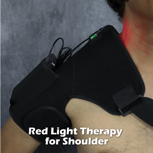
  • Use Red Light Therapy and Pulsed EMF to stimulate cartilage growth.  Check out the video below where I explain how to use them:


Lastly, if you have chronic knee pain and are concerned that knee replacement surgery is a few years down the road, here’s an inspiring article.  It’s a story from a guy who actually experienced natural cartilage regeneration:

Knee Cartilage Repair: How One Patient Proved His Doctors Wrong

The Bottom Line:  First and foremost, to avoid a future of osteoarthritis of the knees or hips and all the limitations it causes, think prevention — diet and lifestyle modification.  Natural cartilage repair and regeneration is still possible, but it is extremely slow and only works if the damage is minimal.   Intervene early, not later.  Go heavy on nutrition that supports cartilage growth because, as this blog post asserts, the cells that make cartilage are still alive and active.  They just need some help.

Unresolved Joint Ligament Pain – Possible Explanations, and How to Treat

sprainSometimes when you injure an ankle, wrist, knee, finger or other joint, the pain doesn’t go away entirely.  A nagging soreness remains, months and even years later, and your joint is not at 100% capacity.

A ligament connects one bone to another; whereas a tendon connects a muscle to a bone.

A sprain refers to an injured ligament; a strain refers to an injured tendon.  When you injure a joint, ligaments and tendons are usually injured together; thus the common term used by doctors for this injury:  “sprain-strain.”

 The main reason why ligaments and tendons take a long time to heal compared to muscle and skin is that they don’t have a direct blood supply.   They rely on absorption of extracellular fluids, much like spinal discs.

Ligaments and tendons, a class of connective tissue are mostly comprised of a collagenous matrix secreted by special cells called fibroblasts and chondroblasts, and it takes considerable time to make this matrix.  That is why when a pro athlete significantly injures a ligament, it is usually a “season ending” injury due to the long time it takes to heal.

 Sprains and strains are graded I-IV.  Grade I is a minor sprain-strain, where just a few fibers are torn.  Grade II sprains and strains are partial tears, where the tear doesn’t go all the way through the ligament or tendon.  Grade III is a complete tear, where it separates, and Grade IV is when it comes off a bone.

Possible Causes of Chronic Ligament Pain

 If you injured a joint and it is still painful after a month, and you notice instability (the joint moves more than it should) then you probably sustained a Grade III or IV sprain-strain.  It is the instability that stresses the rest of the joints ligaments and tendons and prevents complete healing.  Most people get the ligament/tendon reattached surgically; some leave it alone which is not a good idea because the instability will accelerate joint degeneration resulting in more pain and more loss of function down the road.

 If you sustained a Grade I or II strain but still feel pain, it could be that you are continually aggravating it, or on the opposite side, keeping it too immobile causing it to atrophy and delay healing.

 Another possibility of chronic ligament and tendon pain is scar tissue adhesions.  When ligaments, tendons, fascia and muscle are injured, the body lays down collagen scar tissue, which is less organized than normal collagen fibers and tends to bunch up and calcify.  It can also stick to adjacent tendons and interfere with their movement, causing pain.

How to Treat a Recent Sprain Strain so It Doesn’t Become Chronic

Unless you have a Grade III or IV sprain-strain, which warrants a visit to the orthopedic doctor, the protocol upon injuring your joint is to rest it, ice it (20 minutes every 2 waking hours for a few days), compress/support it with tape or a brace, and try to keep it elevated to reduce edema.

  • Apply a comfrey root-based ointment to your ligament daily, such as Kytta Salbe, Dr. Christopher’s Formula or Burt’s Bees Res-Q.  Comfrey is a plant that has been used for a variety of ailments for centuries, especially injuries and pain.  It contains allantoin, a cell proliferant that speeds up the natural replacement of body cells.
  • Apply red-light therapy to your ligament three times a day.  Red light is known to increase cellular ATP (energy) production, which enhances healing.
  • Do myofascial therapy using a myobar or Edge tool.  Using long strokes along the length of the tendon, press down firmly but not too hard.  When you feel the small bumps of scar tissue, use shorter, firmer strokes to soften them up.
  • Do resistance exercises to put a controlled load on the affected tendon.  A 5 lb. dumbbell is good for wrist and shoulder problems; if it’s your knee or ankle ligament, use an ankle weight.  Move your joint in all directions with the weight.  This stimulates the fibroblast and chondroblast cells to make more collagen matrix in the ligament.

 Watch this video to see how I use Red Light therapy to accelerate tissue healing:


Lifestyle Modification for Stronger Joint Ligaments

  • Avoid smoking (if you do).  Smoking interferes with healing of any kind.
  • Eat bone broth soup weekly, made with animal joints and bones, and eat all the cartilage and tendons as well.  This provides building blocks for connective tissue.  Pork and beef joints, chicken and turkey carcasses, and fish (salmon spine and heads) work well.
  • Make sure to include onions, garlic and shallots in your diet.  Alliums are high in sulfur, which is needed for protein (collagen) synthesis.
  • Take supplements for joint health such as MSM, collagen and NEM- natural egg membrane, as well as anti-oxidant supplements such as astaxanthin, cherry juice and Vitamin C.
  • Lift weights regularly.  When you put loads on your ligaments and tendons, the cells sense the load and automatically secrete more collagen, making them thicker and stronger.  Use 5-10 pound dumbells to strengthen your wrists by holding the weight in front of you, palm facing down, then extending just your wrist to lift the weight.  This strengthens the tendons at your elbows (protects agains epicondylitis/ tennis elbow).  Read this post on how to strengthent your joints, which goes into more detail.


DOMS: That Nagging Soreness You Get Two Days After Rigorous Exercise

small-runners legs maleIf long distance running, long hikes and/or doing leg presses or squats are things you do often, you likely notice soreness and stiffness in your legs.  I mean intense soreness where you actually feel deep pain in your muscles, and your legs feel like solid lead (the metal), making walking a chore. This condition is called Delayed Onset Muscle Soreness, or DOMS for short.

The delayed nature of muscle pain after hard exercise is due to the time it takes for offending biochemicals to build up to levels that are irritating to sensory nerves surrounding your muscles. These biochemicals are creatine kinase or creatine phosphokinase (CPK), hydroxyproline and lactate dehydrogenase.  They are normally released in response to muscle breakdown, which occurs with rigorous exercise.  This process is called catabolism.  It used to be thought that lactic acid was the culprit, but further studies doubted that association, as lactic acid is a normal byproduct of anaerobic metabolism and is quickly cleared out as it forms.

The prospect of being sore all over can discourage exercise, which you don’t want to do as exercise improves numerous health metrics such as blood pressure, insulin sensitivity, lean body mass, aerobic capacity and stronger bones and joints.  Instead of being discouraged from exercise, take steps to minimize DOMS and manage it so that it isn’t a problem.

Self Treatment for Delayed Onset Muscle Soreness

1. Immediately after your exercise, fill your bathtub with cold water and soak in it for 20-30 minutes. This dampens the low-grade inflammatory reaction occurring in your muscles from arduous exercise. If your water isn’t cool enough, add some ice cubes.

2. Re-hydrate your body with a quart of coconut water.

3. When you feel DOMS approaching (about 12-18 hours after your activity), soak in cold water again and then follow it up with a warm Epsom salt bath, with some boswellia and lavender essential oils added in.

4. Follow your bath with a 20 minute infrared sauna or red light therapy session.

5.  Use compression boots to help drive stagnant fluids in your legs back into your circulation.  These are great to use after a leg workout even if you don’t get DOMS.

Do not aggressively stretch your legs during DOMS, just wait for the pain to go down, and then do light stretches.

7.  If you want the soreness to go away even faster,  apply Pulsed EMF to your legs after your event, to minimize the effects of DOMS.  PulsedEMF uses low frequency magnetic fields which normalizes muscle cell membrane charges, which restores membrane permeability and allows the cells to return to equilibrium faster.  One of the effects of normalized cell membrane charge is reduced inflammation and increased ATP/ energy production.  Watch the video below where I explain how this works.

Recommended Lifestyle Changes

1. Exercise regularly to keep your muscles toned and conditioned. Engage in short distance running, aerobic exercises, and functional exercises.

2. Consume complete protein six hours and two hours before your activity. I recommend a whey protein or pea protein smoothie (blend with almond milk or coconut milk). Throw in a raw egg as well.  Whey and egg protein have high bioavailability compared to meat, and therefore can be assimilated easier for your body’s use.

3. Take Curcumin and Astaxanthin supplements. These are potent anti-oxidants that can help your body neutralize free radicals generated by exercise. Make sure to take with a meal that contains some fat, as curcumin is not water soluble.

4. Warm up for ten minutes before you do the extreme stuff: stretches, squats, jumps, running in place.


How to Reduce or Eliminate Tension Headaches

HEADACHETension headaches are common to millions of people.  While most cases are not debilitating (enough to incapacitate the sufferer), they still can impact one’s ability to perform and produce.

The cause of headache pain is difficult to pinpoint as there are currently no diagnostic tests that can provide a definitive answer.  But there are definite, known triggers of headaches including certain food allergies, food additives, chemical fumes and noxious visual stimuli.

With careful investigation, one can trace the cause of his or her headaches and take steps to minimize their occurrence or eliminate them entirely.

Those who get tension headaches describe a sensation of a tight band wrapping around the head, constricting it.  There is pressure around the temples and behind the eyes, or in the back of the head and neck but the pain can be anywhere in the head area.

The onset is usually slow, but is definitely noticeable once it starts.  You’ll feel a gradual tightening of muscles (although it may not be the muscles, but a nerve or vascular sensation mimicking tight muscles) around the head especially forehead; and sometimes back of neck.  When it is at its peak, you feel like closing your eyes and stopping what you’re doing.  At this point, most people will take an aspirin or Ibuprofen.

Aspirin works by blocking an enzyme called COX (cyclo-oxygenase) that is used form a substance called prostaglandins, which amplify pain signals and help initiate inflammation.  Reduce pain signals and reduce inflammation, and you can reduce the pain from headaches.  But the problem with aspirin is that it can make your stomach prone to bleeding, as prostaglandins help protect the lining of stomach from stomach acid.

Ibuprofen is a non-steroidal anti-inflammtory drug that doesn’t have the risk of stomach bleeding like aspirin does, but is known to cause damage to the kidneys in high doses or frequent use.

If you are prone to getting tension headaches i.e. get them regularly, then chances are you are doing something on a regular basis that is triggering them.  Your goal is to identify these triggers and eliminate them.

Here are the steps:

  1. Take a food allergy test.  You can get a home food allergy test kit to determine if you are allergic to a particular food.  “Allergy” in this sense does not only refer to sinus-related symptoms like sneezing and a runny nose.  It refers to an immune response to eating a certain type of food that may manifest as headaches, GI upset, fatigue, joint pain and a wide variety of other symptoms.  Once you identify one or more foods that trigger a response, eliminate them immediately.
  2. Keep a daily log of your activities— places you go/things you do regularly during your week and potential headache triggers that may be in those  environments.  Things to look for:  heavy machinery, office machinery, paints, chemical fumes, food, drinks, and high energy electronics.  See if your headaches coincide with visiting any of these environments.
  3. If you work with a computer/laptop, try reducing the screen brightness.  Try your best to unplug two hours minimum before bed time.
  4. Tension headaches can be caused by periods of continuous concentration.   If you have a job that requires this, have you heard about the Pomodoro Technique?  The Pomodoro Technique is an innovative time management system that basically advocates breaking up your activities into 2o minute chunks, separated by short rest periods.   This enables your brain to recharge itself for more work. More info can be found here.
  5. If you tend to hyperventilate, or have a low respiratory rate, you may be building up higher than optimal levels of blood CO2.  This causes blood vessels to constrict, which can trigger headaches.  To solve this, practice Butkeyo breathing.
  6. Lastly, regular tension headaches may be caused by neck bones being out of place.  The neck bones, or cervical vertebrae can easily shift out of place from accidents and poor postural habits, changing the biomechanics of your cervical spine (neck).  This can cause the neck alignment to straighten or reverse, as opposed to having a nice arc for balancing.  One or several vertebrae may be rotated or tilted to one side, causing muscle strain and abnormal pressure to the joint surfaces (see if you have a head tilt).   An abnormally aligned neck can also irritate the spinal cord and/or spinal nerves that exit down to the arms and back muscles.  All these factors can lead to tension headaches, among other things.  Read my previous post on how to self-treat neck problems here.

If your neck feels out of place; i.e. you can’t turn or bend it fully and/or you feel neck tension, then consider getting checked by an experience chiropractor.  Chiropractic adjustments can help restore movement and alignment to your neck and potentially reduce or eliminate your headaches, if the cause is due to abnormal neck alignment.


What Causes Radiating Arm Pain?

Just like how sciatica/ leg pain is usually caused by a bulging disc in the lower back or by a muscle pinching the nerve, arm pain and/or numbness & tingling is usually caused by a bulging disc in the neck or by a muscle pinching a cervical (neck) nerve root or plexus.

The nerve roots that come out from between your cervical vertebrae converge to form three main nerves that service the arm: the ulnar, median and radial nerves. If any of these nerve roots are pressed by a bulging disc, a bone spur or thickened ligament, it usually causes radiating (traveling) pain from the neck down the arm; usually all the way down to the fingertips.

The specific area of pain/tingling depends on which nerve root is being pinched. In fact, that is how doctors diagnose the precise problem area. For example, numbness down the medial (inner) side of the forearm down to the pinkie and ring finger can be produced by compression of the C8 & T1 nerve roots. Numbness in the outer forearm, thumb and first two fingers is associated with the C6 & C7 nerve roots.

arm dermatomes

Radiating arm pain can also be caused by thoracic outlet syndrome, where muscles near the neck and upper shoulder pinch the nerve plexus (where the roots converge). This will be next week’s topic.

Self Treatment for Radiating Arm Pain

If you have significant neck pain accompanying your arm symptoms, then it is likely you have a bulging disc in your neck. It feels like a focused, sharp pain deep inside the neck on one side. Massage does not help this kind of pain.

Most mild to moderate cases of cervical disc bulges resolve with exercises and manual therapy. The severe cases usually require surgery to remove the disc portion that is pressing against the nerve. If you have a severe case, get a consultation from a spinal surgeon and a second opinion. If done in time, surgery can resolve the arm pain, but if the entire disc is removed the surgeon will fuse the vertebrae above and below the disc which will reduce your neck range of motion somewhat.

If the disc bulge or offending structure is allowed to compress the nerve root for an extended period it may result in permanent injury to the nerve. This means after surgically removing the bulge, you still may have numbness down the arm. This is why, for nerve compression conditions, time is of the essence.

Non-surgical candidates can do exercises to reduce the bulge size. For typical posterior bulges (bulges that protrude towards the back and one side of the vertebral body) try this: while standing, use your posterior neck muscles to pull your neck straight back, as far as you can; hold for 3 seconds. It may feel uncomfortable if your disc bulge is acute. Keep your chin tucked in so that the top of your head is level. Do eight times, twice a day for a couple of weeks; note changes in your neck pain and arm pain. Discontinue if it aggravates your condition. This movement gently presses the backs of the vertebral bodies together, which pumps the disc bulge back to center.

If the exercises help but you hit a plateau, try side bending your neck towards the side of the pain, very slowly; repeat six times. Again, note changes; discontinue if it aggravates the pain.

Recommended Lifestyle Changes

Forward bending of the neck and anterior weight bearing of the head (forward head posture) tends to make the cervical discs more vulnerable to bulging because in this position the vertebrae press the front part of the discs, pushing the jelly center (nucleus) towards the back.

Axial forces (straight down through the spine) to the neck can also make disc bulges worse. Any activity that involves jumping creates axial forces — running, basketball, gymnastics, mountain bike riding, sky diving, etc. It’s not a concern unless you do it frequently.

If you have a bulging disc in your neck with arm pain, here are some suggested lifestyle changes:

  • Use a contoured neck pillow and sleep on your back.
  • Work on improving your posture: eliminate forward head posture.
  • Get a standing desk if your job requires a lot of sitting– it’s better for your back and neck.
  • Use the Cervical PosturePump device to hydrate your cervical discs
  • Strengthen your neck muscles so they offer more support to your neck.
  • Avoid excessive jumping. If you like running, consider getting Z-coil or Gravity Defyer shoes (see below).

Treatment Accessories to Reduce Arm Pain from Disc Bulge

posturepumpPosturePump Disc Rehydrator

This device uses specially designed air bladders, inflated by a hand pump to spread apart and extend neck vertebrae. This expands the discs, drawing in fluids and nutrients and also stretches the neck into its normal, ideal curvature.


zcoil2Anti-Shock Specialty Shoes

These specially designed shoes have powerful springs in the heel that significantly dampen the forces generated from running. Less shock to your feet, ankles, knees, hips, low back and neck.


standing_deskVari-Desk Height Adjustable Portable Desk

Place this lightweight desk on your traditional sit-down desk and switch its height between standing and sitting in less than five seconds. Choose to stand for as long as you like, then switch back– great for easing into standing while working, if you’ve been a desk sitter for many years.

How to Self Treat Sciatica- Radiating Nerve Pain in Buttock and Leg

Most cases of sciatica involve a bulging disc in the low back pressing on the S1 nerve root, left or right side; less commonly on both sides. The S1 nerve root is one of several that form the sciatic nerve, the largest/ thickest nerve in the body that controls muscle contraction and sensation in the legs.

The S1 nerve root may also be pinched by a thickened spinal ligament or an osteophyte (bone spur) in the foramen (hole) where the nerve comes out of the spine (notice the left vertebrae in the image below demonstrating this).

verteb

Sciatica can also arise from tight muscles in the buttock (hip rotator muscles) squeezing it. If you had a fall or injury that misaligned your sacrum, lumbar spine or hip joint it could throw those butt (gluteal) muscles out of balance, causing them to scissor the sciatic nerve where it passes between these muscles.

pirifomis

Lastly, the worse kind of sciatica is from spinal stenosis. This is when the central spinal canal in the lumbar spine narrows, pinching the nerves that lead to the legs. The canal narrows due to degenerative joint disease in the lumbar spine. Some people in this group walk in a crouched forward position, because it seems to provide relief.

ss

Treating Sciatica Yourself

If you have some low back pain along with sciatica, or a history of it, chances are your sciatica is due to a bulging disc.  The strategy then is to reduce the bulging disc; i.e. decrease its size so that it doesn’t pinch the nerve.
Lumbar extension exercises at various angles can be helpful in reducing bulging lumbar discs.  The key is to find the right angle, as all disc bulges don’t behave the same in response to certain movements.  The correct movement causes the lumbar vertebrae to “pinch” the bulge back to center.   Watch this video where I demonstrate how to do this.

You can also use the PosturePump.  This is a device that extends the lumbar spine in the non-weight bearing position (while lying down).

If your sciatica is caused by pinching of the sciatic nerve in the buttock, try stretching your gluteal muscles (piriformis) muscle to lengthen it.  There are several ways to do it.  One is to cross your legs while sitting (bad leg on top), clasp your hand over the top knee and bend forward as far as you can go (may be difficult if you have a large belly).  Hold for about 15 seconds; relax.  Repeat 6 times twice a day.

You can use a power massager like the Max2 Percussion massager to “shake” the spasm out of the piriformis muscle.  Watch this video where I demonstrate this technique.

If you have canal stenosis, and your symptoms are unbearable, get a consultation from a spinal surgeon on your options.

Sciatica often affects those who sit frequently, for long hours.  Sitting places a lot of pressure on the discs and promotes bad posture.  It also places pressure on the sciatic nerve, especially if you don’t have much fat in the buttocks for cushioning and if you have a hard chair.  Wearing a thick wallet in your back pocket can cause more pressure to the nerve.

One of the best ways, in my opinion, to reduce the bad effects of sitting is to use a stand up desk.  Many employers will pay for this, since studies are coming out showing how prolonged sitting damages health over time.  The VariDesk  is an affordable option for a standing desk.

standing_desk

Other considerations

-Get good shoes with arch support.  This alone may ease your sciatica.
-Strengthen your low back, hip and butt muscles by doing kettlebell swings.
-Seek out an Active Release Technique (ART) practitioner.  This is a special massage designed to normalize muscle contraction and joint movement.

If you have a history of injury involving your low back and/or pelvis, get checked by a chiropractor.  Chiropractic adjustments can re- position the area, removing abnormal pressure to muscles and nerves. 

How Neck Pain Develops, and How to Manage It

neck-pain2In terms of the most prevalent types of spinal pain affecting the general population, neck pain closely trails lower back pain.

For the low back (lumbar spine) it’s easy to see why pain in this area is common:  the lumbar spine bears the weight of your upper body in the standing and sitting positions and is a fulcrum point of body movement, so it is a workhorse that gets quite a beating.

But the neck (cervical spine), while smaller than the lumbar spine and less burdened by body weight (just the weight of the head) has certain characteristics about it that make it prone to pain as well.

Here’s a breakdown of those unique characteristics:

  1. The cervical spine is comprised of seven vertebrae. The first two under the skull are the only set of vertebrae in your spine that does not have a connecting disc. This enables greater range of motion compared to the thoracic and lumbar spine regions.
  2. More movement comes at a cost of less strength. The cervical spine therefore cannot bear forces as well as the rest of the spine and is more vulnerable to injury/ mechanical problems (whiplash, sleeping on a bad pillow, etc).
  3. Forces that act on the cervical spine include gravity, repetitive axial forces from walking running and jumping; abnormal static posture/ sleep positions (stomach sleepers) and trauma from car accidents, falls and sudden jolts.
  4. Structures in the neck affected by these forces are the facet joint surfaces in the rear of the cervical spine; the uncinate joints to the sides of the cervical spine; cervical discs, spinal cord, nerve roots, muscles, fascia, and even esophagus and throat.
  5. Osteophytes, or spondylosis occurs when projections of bone develop usually from abnormal movement over time. Problems occur when these bony protrusions encroach on nerve tissue.

If you have neck pain, chances are it is related to mechanical dysfunction.  But you must also be aware of other conditions that can generate pain and/or tenderness in the neck, and rule these out with your physician if the causation/source of your pain is not clear.

Causes of Neck Pain

Red flags (requiring immediate attention) include:

  • fracture
  • lymphoma
  • bone tumor
  • spinal cord tumor (syringomyelia)
  • onset of rheumatoid arthritis
  • esophageal varices
  • vascular disease (atherosclerosis)

Viral infections are sometimes associated with stiff and deep aching pain in the neck

Primary hyperkalemic paralysis—a rare condition usually triggered by extreme exercise, characterized by high or low levels of potassium, an electrolyte involved in muscle contraction.   Causes severe pain and spasm initially, followed by muscle atrophy and loss of tone and contraction.  The condition usually resolves after a few weeks.  It can occur in the neck, and other muscle groups as well.

Neck Pain of Musculoskeletal Origin

  • Myofascial pain related to previous trauma.  Trigger points and scar tissue are characteristics of MP.
  • Sprains and strains from injury (whiplash, direct trauma)
  • Generalized muscle pain from muscle tension, fatigue or spasm related to poor ergonomics, bad posture and often stress
  • Spondylosis, osteophytes (osteoarthritis related to degenerative disc disease)
  • Spinal stenosis – the narrowing of passageways where nerves pass
  • Disc bulge or herniation
  • Cervical radiculopathy (compressed cervical nerve roots from spondylosis or disc bulge)

Living in a modern society where sitting at a desk looking at a computer monitor in front of you or work papers down in front of you is the origin of many cases of neck pain.

This unnatural position  of craning your neck forward while seated, over time, degrades good posture.   It is unnatural because the human body is optimally designed and meant to stand and walk.

bad_posture

Exercises to Reduce Neck Pain of Musculoskeletal Origin

First, determine if you have a muscle imbalance, where your anterior (front side) neck and trunk muscles are overpowering your posterior (back side) neck and upper back muscles, pulling your head forward of the spine.  Do a posture check, visualizing plumb lines from front and side.   This abnormal posture is referred to as Anterior Weight Bearing of the Head (AWB).    Another term for this is Forward Head Posture (FHP).

If you have AWB, the force to your cervical spine (and muscles) more than doubles compared to if your head’s center of gravity was directly over your torso/ axis of spine.  This is because the weight of your head and the forward angle of the neck in AWB combine to create a moment force.

To get an appreciation of this, imagine holding an eight-pound shot put directly over your head, arm extended.  It should be fairly easy to do.  Then, while still holding it above your head, move the shot put just 5 degrees forward, simulating AWB of the head.  Your arm muscles will quickly fatigue and even develop pain.

The increased forces to your neck  in AWB of the head  get absorbed by your neck muscles, your discs and vertebrae.

Use the diagram below to evaluate your posture (you may need help from another person to view your posture from the side).

posture

Ideal human posture, viewed from front and side. From the front, a line bisecting the center of the body should have equal amount of body mass on each side (symmetrical sides). From the side, the gravity line should pass through the ear, center of shoulder, hip, knee and slightly behind ankle.

If you have AWB/FHP, engage in exercises to counter and correct AWB/FHP, with the goal being to reduce the amount of it:

Neck extension exercises – While lying face down on a mat, arms to side, contract your posterior neck muscles and lift your head straight up without arching it, as high as you can.  Hold for four seconds; repeat 12 times 3x/ day.

Neck mirror image posture correction exercises – If your neck tilts to one side abnormally, stand with the opposite side shoulder contacting a wall.  Using your neck muscles opposite the side of abnormal tilt, pull your head sideways towards the wall without bending your neck (keep your head level during the exercise).  Hold for four seconds, repeat 12 times 3x/day until your neck is centered.  Repeat as needed.

Wall angels.  See this video of me demonstrating this exercise.

Isometric strengthening exercises for neck – Find a small child’s inflatable ball and place it against a bare wall, head level.  Using different parts of your head each time (forehead, side of head, crown of head), press the ball against the wall and hold for 10 seconds.  This isometrically contracts your neck muscles, strengthening them.

Rolled towel exercise to improve curvature-  Roll up a towel to about a 5″ diameter, or better, buy a cylindrical neck pillow.  Place it underneath your neck as you lie on your back.  Arch your neck over the pillow and press the crown of your head onto the floor; hold for five seconds and repeat ten times.  Next, with the roll still under your neck turn your neck as far as you can to the left, then right, five times for each side.

Use the Posture Pump Disc Rehydrator device

Thoracic outlet exercises: shoulder circles, corner stretches

Quadriceps stretch  Tight or shortened quadriceps muscles (your lap muscles) can rotate your pelvis forward causing your upper back to lean forward which can strain your neck as it tries to correct.  To stretch, while standing bend your leg at the knee straight up keeping your knee pointed down.  Grab your instep with the same side hand (balance yourself, as you will be standing on one leg) and pull it straight up.  You should feel a stretch to your quadriceps muscle.  Hold for ten seconds; switch sides.  Repeat five times each side.

Core exercises – It’s important to have a strong core to support the lumbar spine properly, which fosters better posture overall.  Do crunches, medicine ball exercises and planks to strengthen your core.

If you have any history of previous neck trauma such as sports injuries or repetitive movements, car accidents, falls etc., or spend a lot of time at a desk in a static neck posture, it’s possible that one or several of your neck vertebrae have locked together or have lost some movement which can have the effect of perpetuating discomfort. Chiropractic adjustments can be helpful in restoring motion to these segments and when combined with rehabilitative exercises, can usually resolve cases of general neck pain.

Lastly, a great way to discourage formation of AWB and encourage good posture is using a standing desk.  The VariDesk is a quick and affordable solution to standing while working.  Lower it, and you’ve got a traditional sit down desk; raise it (takes 5 seconds) and now you’ve got a standing desk.  Alternate between sitting and standing throughout your day and notice your neck and back pain and stiffness improve!

standing_desk

The VariDesk can dramatically reduce low back and neck pain, and upper shoulder tightness.

Summary:

If you have neck pain and you aren’t certain of the causation/origin, see your doctor and rule out red flags.

X-rays are helpful in assessing the state of your cervical spine (alignment, disc spacing, bone density, abnormalities, level of decay).

Countering anterior weight bearing/ forward head posture, strengthening the neck muscles, improving your cervical curvature, stretching your leg muscles and strengthening your core will improve your posture and reduce strain and pain to your neck.

Why Skinny Thin People Can Still Have Low Back Pain

It’s probably a safe bet to assume that people with chronic lower back pain are more likely than not to be overweight or obese.  Although there are a few exceptions related to genetic disorders and medical conditions–thyroid disease, Cushing’s syndrome, depression– those who are overweight got that way because they are less physically active and do not eat as healthy as those who are not overweight; i.e. they consume more calories on average in their diet.   This is attributed to mindset, which is a major contributor to, if not actual origin of most types of chronic disease (heart disease, cancer, diabetes, high blood pressure).   With excess weight comes excess pressure to the weight bearing joints of the low back, hips, knees, ankles and feet; hence the association between back pain and overweight individuals.

But what about those folks who are normal weight, or even under weight and have terrible episodes of low back pain?

It seems highly unlikely, but it does happen.  After all, how can a skinny person who doesn’t have much fat and muscle to carry around develop low back pain?

If you are thin and have recurring lower back pain, here are some possible explanations:


Bad genes

Ongoing research is finding a connection between certain gene markers and lumbar disc degeneration.   If you possess such markers in your genetic profile, you are more vulnerable to developing degenerated discs, which are a common source of lower back pain.

The good news is that such bad gene markers need to be activated in order to do damage.  You may be able to delay, or prevent this activation by practicing a healthy lifestyle– eat naturally occurring foods with copious amounts of anti-oxidant and nutrient-rich green, leafy vegetables; regular, moderate exercise, adequate rest/ deep sleep; minimizing toxins (alcohol, sugar, tobacco, pollution, chemicals in cosmetics and food additives); and engaging in socially rewarding activities.  The opposite behaviors are the very things that can trigger activation (up-regulating) of bad gene markers, initiating the sequence of events that eventually will manifest the disorder– smoking, drinking, junk food, lack of exercise, pollution and so on.

You are sedentary most of the day

You don’t have to be overweight to be sedentary.  If you have a high metabolism or simply don’t gain weight despite eating junk food, big lunches and late night snacks, don’t celebrate–  you may be skinny but unhealthy in several health metrics like strength, energy and stamina; cardiovascular endurance and insulin sensitivity.

Sedentary people sit more than they stand in a day and stay relatively motionless (TV, internet jockeys) and don’t exercise or do physically demanding work.    A sedentary lifestyle leads to muscle atrophy in the legs, pelvis (hips, buttocks), abdominal muscles and spinal (postural) muscles.   Those muscles groups, because they aren’t firing together often, lose coordination with one another.  The autonomic part of the brain “forgets” how to make them contract properly, in proper synchronicity, during every day movements such as bending and twisting of the torso; lifting objects from a low position to a higher one and rising off a chair.   As a result, the lower back does not get proper support, opening it up to injury.

Sedentary individuals are prone to experience an acute low back injury when trying to move something heavy or suddenly engaging in physically demanding activity; or their low back pain may develop from insufficient support to the lumbar vertebrae, causing weak back muscles to strain and joint surfaces to get overly taxed.

Previous injury or history of cumulative force trauma to your spine

If you played a sport or recreational activity when you were younger that involved jumping and landing, you may have predisposed yourself to disc degeneration with all the repetitive trauma to your spine and weight bearing joints.  Sports that fall into this category are gymnastics, basketball, football and volleyball.   Motocross, parachuting and martial arts are other activities that can result in cumulative force trauma to the spine.  Such forces over time pound the L4/5 and L5/S1 discs and may even damage the vertebral end plates of the vertebrae above and below.  When this happens, that area calcifies and nutrient absorption from the tiny capillaries in the end plates into the disc is reduced.  As a result, degenerative joint disease accelerates.  The disc thins and forms painful tears and/or bulges.

It’s also possible that the cumulative force trauma caused a pars stress fracture, or spondylolysis that is making your L4 or L5 vertebrae unstable, where it shears back and forth during bending of the waist, irritating ligaments and nerves.

If the following signs and symptoms apply to your particular low back pain, there is a good chance you have a pars fracture and/or instability of your L4 or L5 vertebrae:

  • adolescent athlete
  • low back pain predominantly on just one side of the lower back
  • started as mild pain; worsens with running and jumping
  • feels the worst when arching backward, twisting the waist or straightening your back from bending
  • gets worse with sports or heavy work, and better with rest

pars stress fracture xray

If jumping or contact sports are in your history, get a motion x-ray study (x-ray views taken in lumbar flexion, neutral, and extension in the weight bearing position), or video fluoroscopy study.   The x-ray series will reveal if one segment is moving abnormally relative to the ones above and below.  If diagnosed, the standard approach to treating spondylolysis is to modify your movements to reduce the shearing effects; strengthen the core muscles so that they offer more stability to the lumbar spine; lumbar bracing and perhaps shoe orthotic inserts.

Unfortunately, pars fractures do not usually heal due to the delay in discovering them, and the difficulty in bracing them.  In some cases, the gap is closed by fibrous tissue the body lays down, which offers some stability.

Vegetarian Diet

Your vegetarian diet (if that’s the case) could be contributing to back pain.  Vegetarians are more susceptible to having Vitamin B12 deficiency, since Vitamin B12 is only found in animal sources.  Vitamin B12 is the “energy” vitamin and plays a big role in a number of important biological pathways.  Studies show that low levels are associated with  ataxia (shaky movements and unsteady gait), muscle weakness, spastic muscles, incontinence, low blood pressure and vision problems.

Vegetarians may not be getting enough sulfur in their diets.   Your body needs sulfur to build strong muscles, bones and cartilage, among other things.  Sulfur is also believed to be protective against glycation— the harmful reaction where excess glucose combines with proteins in tissues, effectively denaturing them (rendering them useless).   While plants like onions, leeks and asparagus contain sulfur, animal protein is the most complete form.

Lastly, vegetarians may not be getting enough cholesterol in their diets.  Cholesterol is needed for healthy cell membranes, proper nerve function and synthesizing cortisol and sex hormones.

A great meal to counter-act all these deficiencies is bone broth soup.  Packed with calcium, collagen, elastin, chondroitin, sulfur and more, bone broth soup is great for your skin, hair, nails, connective tissue, nerves bones and muscles.   Not too many meals can compare, nutrition-wise to bone broth soup with spinach!

Osteopenia

If you are a female whose mother has osteoporosis, you may be carrying the gene.  Osteopenia is the loss of bone mass and occurs when your body does not replace calcium as fast as it resorbs it (releases it from your bones into the blood).  It may be related to low Vitamin D levels, thyroid disorders, estrogen deficiency, hysterectomy or other condition which would cause bone to lose calcium.

Osteopenia leads to osteoporosis, where the cancellous or spongy, inner part of a bone has lost much of its density, thereby weakening the bone.  Osteoporosis of the spine leads to a humped/ stooped posture as the vertebral segments shrink, and fragile bones highly susceptible to fracture.   Spontaneous vertebral body fractures are common in osteoporosis and an often overlooked cause of back pain.

If you suspect osteopenia, I would advise getting your Vitamin D levels checked.  Vitamin D is necessary for your body to absorb calcium from you diet, in your intestines.  If you are low, make it a point to expose your body to the sun 4 hours a day.  The UV rays in sunlight initiate the synthesis of Vitamin D3 from cholesterol present in your skin.  Then it is converted to another form in the liver (calcidiol) and finally to the active form (calcitriol) in the kidneys.  So your liver and kidneys need to be healthy– avoid alcohol, tobacco, drugs, unnecessary medications.

Also, take high doses of Vitamin D supplements.  Based on current research, consuming 1,000–4,000 IU (25–100 mcg) of vitamin D daily should be ideal for most people to reach healthy vitamin D blood levels.  This may seem high, but know that much of what you take does not get absorbed; around 30%.

Lastly, did you know that Pulsed Electromagnetic Field Therapy (PEMF) can increase bone density?  PEMF is the external application of compatible electromagnetic fields to the body to augment the body’s naturally occurring fields, which helps cells and tissue function more optimally.  PEMF is often used to heal non-union fractures, as well as help reduce inflammation and pain.

Bio Balance Pulsed EMF

You can read more about how Pulsed EMF works here.

The bottom line, thin people get back pain, too.   If you are normal weight and are experiencing lower back pain, look into these possible causes and take action.  All of the above factors can be positively affected by making changes in your lifestyle.

If you were diagnosed with degenerative disc disease, check out the recommended home therapy devices in our Amazon store:

Try using the PosturePump

posturepump elliptical spine trainer


This device decompresses the lumbar discs so that they can rehydrate and heal faster.   Simply place it under your low back as you lie on the floor, and pump up the air bladders to your tolerance.  Let the PosturePump spread your discs apart in this position for 10-15 minutes, twice a day.  Your pain should face as more space is created by thickened discs.

Fight back against hard to avoid sedentary behavior!  If you are one of the millions of people who must sit hours and hours behind a desk at work, consider getting a standing desk.  Standing relieves some pressure to your low back by transferring some of it to your legs and by encouraging a lordotic (inward curved) low back.  Here is a low-cost solution:

Vari-Desk Stand Up Desk

standing_desk

Top Five Low Back Pain Relief Strategies

djdWhen your low back hurts, you know the next couple of days aren’t going to be fun.  Your low back, or lumbar spine is like the foundation of a house:  it bears and balances all the weight above it, so if there are mechanical problems, the whole structure is affected.  In fact, some cases of low back pain cause obvious tilting of the upper body to one side when standing, due to the structural imbalance.

There are countless studies on low back pain– how it develops; what causes the pain; who is at most risk for developing low back pain; how long it lasts and so on.  For “non-specific” low back pain where there is no injury event, it is unclear which structures are generating the pain.   It may be the back muscles, a vertebral ligament; a disc, nerve root, sacroiliac joint; or even referred pain from an organ.  This makes treatment, especially invasive treatment (surgery, injections) challenging and often ineffective.

What is known is that if you have low back pain that continues down your buttock, and perhaps down the back of  your thigh and leg all the way down to your foot, then it is very likely due to a bulging disc in your low back pressing on a nerve root.  If this describes you, don’t give up hope.   I’ve seen cases like this resolve with targeted exercises, spinal adjustments and time.  But I’ve also seen cases like this turn into back surgery cases, where the surgeon goes in and removes part of the disc that is compressing the nerve, and sometimes the entire disc and fusing the adjacent vertebrae.

Non-radiating back pain can be equally bad.  If the back muscles spasm, the pain can be incapacitating.  I’ve seen many cases like this where the patient dropped to the floor, wincing in pain and avoided any type of movement.  Aside from a cortisone injection, there is not much one can do initially for acute back spasm other than ice and rest until the muscles let go; then attempt things like chiropractic adjustments, PT, stretching and modalities like laser therapy.

Then of course, the rare causes of low back pain — cancer, tumors, bone cysts and other diseases; spinal stenosis, referred pain from kidney stones; fractures, and spondylolisthesis (slippage of vertebrae during movement).  These will require diagnostic testing and proper management.

But for the vast majority of low back pain cases, there are things you can do to reduce their effects and reduce their incidence of occurrence.  Here are my Top Five low back pain relief strategies:

Lose weight

(if you are overweight).  This is just basic, common sense.  If you are 30 pounds overweight, it’s like a normal weight person carrying a backpack full of rocks all day.  Excess weight centered around the abdomen changes the center of gravity of your body.  This bends your spine abnormally when you are standing, sitting, and even sleeping; setting the stage for back pain.

The best and fastest way to lose fat weight is to restrict carbohydrates in your diet, practice intermittent fasting, and exercise 30-60 minutes at least 3x/day.  If you have to cut one of these out; I’d say the exercise.  Caloric restriction burns fat cells on a much larger scale than exercising does.  The reason is that simply by being alive, your body, depending on your mass, burns 1500-3500 calories per day.  If you consume less than what you burn, you will lose fat weight.  So, try eliminating bread, pasta, bagged and boxed snacks, sugar and fruit juice from your diet.  Drink only water.  Eat between the hours of 9 am and 4 pm, or 12 pm and 6 pm only.  This gives you an 18 hour fast, where your body will start burning its fat stores.

Stand more

When you sit, the natural inclination is to round your lower back.  You may try to force yourself to sit erect for a few minutes, but after a while, your low back muscles fatigue and allow your lumbar spine to bow out again.  This posture weakens the lumbar spine’s structural integrity because it separates the facet joints (see middle image below), which makes the lumbar spine less sturdy (“unlocks” it).  The angle of the vertebral bodies also apply a backwards force to the disc that can encourage posterior disc bulging or herniation.

bad-spine

The bad health effects of prolonged sitting goes way beyond back pain.  The amount of time a person sits during the day is associated with a higher risk of heart disease, diabetes, cancer, and death, regardless of regular exercise, according to a review study published in the Annals of Internal Medicine.  This is significant, because computers are firmly integrated into the fabric of life– just about every business from farmers to tech giants rely on computers, and as for now, they are mostly used at a desk.  Low back pain cases can only go up if more and more people spend their work hours sitting behind a desk.

A low tech solution to this is to use a standing desk, like the height-adjustable Vari Desk.  The Vari Desk is actually a large monitor stand that sits on a standard desk and can easily switch from normal height to standing height.  This way, you can gradually increase your standing hours, allowing you to build up your tolerance to working while standing.  For example, for the first week you can try standing 30 minutes every two hours; then work up to standing 30 minutes every hour, then up to three hours straight.  Standing activates the erector spinae spinal muscles (which are inactive when sitting, leading to weakness and atrophy); but best of all standing naturally causes you to form the stronger lordotic curve in your low back.  The lordotic curve is the opposite of bowing and is the “strength” position of the lumbar spine as the facet joints in the back interlock, offering stability and protection to the discs.


standing_desk


Do functional exercises

While your back is not hurting, why not strengthen it to keep it that way?  This will decrease the incidence (occurrences) of low back pain.  Functional exercises include squats, medicine ball exercises, planks, kettle bell swings and “bootcamp” style exercises that focus on strengthening the muscle orchestration of all the muscles in the body at the same time, rather than one type of muscles (for example, biceps curls is not a functional exercise).

Do Back extensions

For most people with back pain, back extensions are helpful.  Again, this arcs the low back in the lordotic curve, which can press the disc material back to center.  You can do them standing, or prone (yoga cobra position).  Do about 10 extension exercises twice a day.

Spinal Adjustments

If you have a history of back pain and/or stiffness; trauma to your body such as prior car accidents or previous involvement in gymnastics, football or basketball; and/or a history of prolonged sitting you are a good candidate for periodic spinal adjustments, or spinal manipulation.  Spinal manipulation, the practice of moving segments of the spine using manual (by hand) techniques, can reduce the symptoms of lower back pain.  Chiropractors provide most spinal manipulation in the U.S.  Some physical therapists and osteopaths do it as well.

Chiropractic adjustments are quick, low amplitude-high velocity manipulations to the spine, which differs from “long lever” movements (joint mobilization, passive stretching).   They are done to impart better movement and also to “fire” nerves involved in pain and spinal muscle contraction.  This can reduce pain and muscle spasm and restore proper movement to a joint segment.  When a lumbar joint segment moves better, it improves hydration to the discs and ligaments.  When it is properly aligned, it won’t wear out as fast.

Consistency in approach varies greatly from chiropractor to chiropractor as there are more than one ways to manipulate the spine, and different target goals (pain relief vs. attempting to change curves of the spine, etc.).  Then there are “holistic” chiropractors who promote spinal adjustments for general health maintenance.  I recommend that you find an experienced chiropractor with a good, local reputation who focuses on short term care to relieve pain and can advise you on home care exercises after your pain has subsided.

Below is a video I made that explains the logic behind lumbar spinal adjustments for low back pain:

Integrate these five practices, and I believe that low back pain will not be a factor in your life for a long time to come.

Receive a FREE, 30-Day Plan to Boost Your Health and Eliminate Pain!

Receive a FREE, 30-Day Plan to Boost Your Health and Eliminate Pain!

As a subscriber, you'll also learn the special methods used by experts in human biomechanics to fix body aches and pain the RIGHT way, long term. 

We'll also send you a Free eBook, Concepts of Self-Healing as a way of saying thanks.

Please check your email in 5 minutes to access your Special Report. Make sure to whitelist "newsletter@painandinjurydoctor.com" in your email client (Gmail, Yahoo, Outlook, etc.) so that you don't miss this valuable information. One way is to add this email to your email Contacts.