Updated 3/20/2021
Most people have sprained their ankle at least once in their lifetime, me included. It can hurt like the dickens and for some unfortunate people it can require a visit to an orthopedic surgeon and installation of surgical screws.
Thankfully, the vast majority of sprained ankles can be treated at home, and resolved 100% in time.
Most sprained ankles are inversion sprains. Inversion refers to movement of the foot inwards relative to the ankle; i.e. “rolling inwards” of the foot while contacting the ground while bearing body weight. Ankle sprains can occur when the person steps on or in something that s/he wasn’t expecting; for example a stone or a hole in the ground. They also occur from sports activities. Wearing the wrong kind of athletic shoes increases the chances of spraining an ankle; for example wearing running shoes while playing on a basketball court.

Basic Ankle Anatomy
The ankle is a synovial joint (encased in a ligamentous capsule and lined with a thin layer of tissue called synovium, which produces synovial fluid for lubrication) that connects the lower leg with the foot (as a side note, synovial joints are the type involved in rheumatoid arthritis). The articulating bones of the ankle are the tibia and fibula bones of the leg, and the talus bone of the foot. Directly underneath the talus is the calcaneus, or heel bone which serves as an attachment point for some of your ankle ligaments.
All of these bones are all lined with cartilage, which absorbs shock and reduces friction in the ankle joint. A major weight-bearing joint, the ankle evenly transmits the weight of the body through the bones of the foot.
There are several, small ligaments that connect the foot to the distal, lateral fibula, also called the lateral malleolus, or outer ankle. Likewise, there are small ligaments that connect the foot to the distal, medial tibia, called the medial malleolus or inner ankle. If the foot starts to roll in quickly as in an impending inversion sprain, mechanoreceptors (movement sensing neurons that send positional information to your brain) around the ankle sense the sudden movement and reflexively correct your ankle to avoid tissue injury. Think of those times you almost tripped, but caught yourself; those are your mechanoreceptors in action. If successful, you avoid spraining your ankle (it also helps to have good balance).
But if the mechanics and circumstances of the movement are such that they overwhelm this corrective reflex, the ankle joint will move past its maximum range of motion, which exceeds the ligaments’ load capacity resulting in a sprain injury. Remember, ligaments connect bones to bones while tendons connect muscle to bone, so a sprain is injury of ligaments while a strain is injury of tendons. They often occur together during injuries and are referred to “sprain-strain injuries.”
The primary function of ligaments is to hold bones tightly together while allowing some movement. They do not have the specialized, contractile cells of muscles, but they can stretch (have elasticity) to some degree, due to the arrangement of the triple-helix collagen fibers within the matrix that act like a strong spring.
Below is a microscopic view of a human ligament (stained), showing tightly packed, spring-like collagen fibers. The dark objects are fibroblast cells.

In a sprain, ligaments tear to some degree. Doctors describe the degree of ligament tears in grades, depending on severity:
Grade 1 – MILD. Painful, minimal tearing of ligament fibers. Ligament typically fully heals.
Grade 2 – MODERATE. More painful, significant tearing of ligament fibers. Can result in joint instability.
Grade 3 – SEVERE. Pain, but sometimes not as painful as Grade 1 or 2. Complete rupture of the ligament fibers. If it’s a major ligament, it will require surgical re-attachment.
This sets in motion the body’s inflammatory response, which is a normal part of tissue healing: a cascade of biochemical substances are released and the ankle starts to swell. Maximum swelling is typically reached in about 2-3 days. There may be a purplish color as well that gets progressively darker, which indicates leaking of small blood vessels pooling in the extracellular spaces. Over time, the body clears the pooled blood out, and the color fades.

Sprained ankle showing blood pooling in multiple areas.
Here’s the thing about inflammation: it is necessary for proper healing, but it can overcorrect causing unnecessary pain and prolonged resolution of the injury. This is why doctors advise using an ice pack during the intial stages to control the inflammation as cold constricts blood vessels making them less leaky. Cold also slows down nerve pain signals to the brain.
Self Treating a Sprained Ankle
The first order of business is to do a simple assessment of your injury. This will determine if you need to see a doctor. Note how much your ankle bent: was it a short and quick sprain that your reflexes were able to correct before serious damage occurred? Or, was your sprain such that you felt your foot bend completely inwards and had little control over correcting; and perhaps you felt and/or heard a snap? When you grab your foot and attempt to move it, does it feel like there is no resistance, as though you ruptured all the ligaments, and it isn’t as painful as you would expect?
The second scenario is a Grade 3 sprain and warrants a visit to the doctor; the sooner the better. These are the signs of ligament and possible tendon rupture, and even ankle or foot bone fracture. With this kind of sprain there is increased probability of internal major vessel rupture, too, which can lead to a dangerous blood clot.
The first scenario is a Grade 1 or 2 sprain. While the second scenario warrants a visit to the doctor, the following treatment can be done in both scenarios.
Old school treatment for soft tissue injuries is to use ice for the first 2 days, then heat. But this is 2021, and there are new things available to treat injuries more effectively, which you’ll learn below:
1. Get home, take off your shoe and sock if you are wearing them. You should notice some swelling/ puffiness around your ankle. Put two cups of ice cubes in a Ziplock freezer bag, and place over your ankle. In the meantime, prepare an ice massager by filling a plastic cup with water and sticking a spoon in the middle, holding it in place with masking tape. Put it in your freezer. When frozen, pull it out of the cup by the spoon handle, and ice your ankle with it.
Don’t have the time for this? Then check out this cheap ice massager on Amazon. It’s even better.
The advantage of using an ice massager like this is that you can also use it to compress the swelling at the same time by pressing it into the tissue as you ice, firmly but not too hard. This helps drive out the swelling. Remember, R-I-C-E soft tissue injuries: Rest, Ice, Compress, Elevate. You can also use a plastic ice massager, like the one I’m demonstrating in this video which you can get on Amazon. Do it for 20 minutes every two hours, for the first two days.
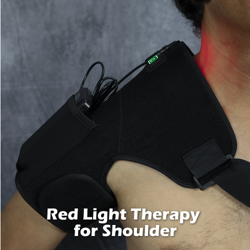
2. Apply Red Light Therapy later in the day, and 3x/day for 10 minutes until it is fully healed.
Red light in the 660nm wavelength range is known to have healing effects via photobiomodulation. Apply it around the lateral malleolus, pressing it firmly but not too hard into the tissue where the swelling is occurring. You should feel the pain go down as you are doing it; it’s that fast.

3. (OPTIONAL) If you want/need to make the pain go away faster and cut healing time in half, apply Pulsed Electromagnetic Field Therapy, or Pulsed EMF/ PEMF to your injured ankle daily until it is healed; 15 minutes, 2-3 times a day. This is an option for you if your job demands frequent walking. A Grade 2 ankle sprain can make walking pretty difficult for about an entire week.
Pulsed EMF is ideal for soft tissue injuries because it energizes cells that need more energy, i.e. cells in injured tissues. PEMF devices emit a low frequency (safe) electromagnetic field that mimics the body’s own naturally occurring fields, augmenting them. Electromagnetic fields drive molecular movement in your body, which is also referred to as cell membrane potential (like voltage in a battery). This boost of energy helps cells carry out their biological functions more efficiently such as nutrient transfer, protein synthesis, and waste removal. It’s like speeding up a video so that it finishes faster, but its the healing you are speeding up!
You can use a Pulsed EMF coil belt device and wrap it around your ankle; or this hand- held Pulsed EMF device, which you can hold over the ankle or use an elastic band to hold it in place for 15 minutes. They aren’t that cheap, but you’ll own equipment to heal injuries or pain you might sustain in the future, so they make a good investment in your health and well- being.

Animation of a fibroblast synthesizing and laying down collagen fibers.
4. Move your ankle. Passively (use your hands) and actively (use your leg/foot muscles) move your ankle starting a few hours after the injury, even if it is a little painful. As your tissues heal, tiny cells called fibroblasts lay down scar tissue and collagen. Moving your ankle through all its ranges of motion stresses the ligaments in the orientation of their fibers, which helps properly align the newly-formed fibers. This is why immobilization (bracing and casting) is not recommended for these types of injuries. However, you can wrap kinesiotape around your ankle with slight pressure to help reduce swelling. Kinesiotape offers support, but also allows movement.

A wobble board is useful for rehabilitating a sprained ankle.
As your ankle pain dissipates, consider using a wobble board to further strengthen/ rehabilitate all the muscles, ligaments and tendons that control ankle movement.
Bottom line, most sprained ankles can be effectively treated at home using the above approach. It is important to ensure proper healing of any joint sprain-strain injury, because you risk re-injuring the same area more easily if the soft tissues heal sub-optimally, such as excessive scar tissue build up and loose ligaments that allow excessive joint movement, which can accelerate arthritis in the joint.

walking “shoe.” It offers the protection of a shoe, while offering the most freedom of movement of your feet.


