It’s been a couple of months since I’ve updated Ask Dr. P. You see, I’m one of those individuals who has multiple interests. We’re living in interesting times, where access to information is becoming easier and easier especially with the arrival of AI, especially ChatGPT, Gemeni, Claude and others.
In fact, I’ve been dabbling with ChatGPT, using it to answer questions about Biology (I teach Biology at the high school level) and I must say, I am very impressed with the results I get.
When the Google search engine came out, it took a couple of years for Google to be universally used. I believe that with ChatGPT, it’s going to take much less time before everyone is used to it, and starts depending on it.
The more you use it, the better you get at it– you’ll get a hang of how to articulate your questions, and a sense of what it can help you with:
an email to someone?
an important letter to a business?
a business proposal?
a college admissions essay?
a recommendation for a European travel itinerary?
and…recommendations on how to heal pain and injury?
The big question on everyone’s mind is, how disruptive will ChatGPT be? Will it enable people to avoid visiting websites like Ask Dr. P to get the information they are looking for?
I think “yes,” ChatGPT, once people are familiar with using it, will at some point become the world’s most visited website. Imagine getting accurate (in most cases; still evolving), concise explanations on how to overcome a problem/ challenge. Will a future version of ChatGPT generate its own videos to accompany its explanations? I think so.
But until then, I will continue to post content on how people can self-manage pain and injuries. I believe that most people still trust humans, and/or connect and learn from them better (as long as they are genuine and qualified) than some online software.
Remember, the means to achieve a pain-free life that are available to you, that don’t include visiting a doctor are:
Diet and Nutrition
Exercise and Stretches
Manual Therapies
Devices and Supports
I produce videos on my YouTube channel in each of these self-help categories.
If you are experiencing chronic aches and pain affecting your back, neck, joints, muscles and tendons then I can teach you how to effectively eliminate or minimize its impact on your life.
My next project is to complete my Ask Dr. P Home Page that indexes all the body parts, and how to relieve or eliminate pain in each of those regions, so please stay tuned.
Imagine waking up one morning, eager to start your day, only to feel a sharp, stabbing pain in the bottom of your heel as soon as your foot touches the floor. It might ease as you move around, but the discomfort often lingers, especially after periods of rest or extended activity. If this sounds familiar, you may be experiencing plantar fasciitis, one of the most common causes of heel pain.
This condition affects millions of people worldwide and can disrupt daily life. In this article, we’ll explore what plantar fasciitis is, its causes, risk factors, treatments, and what to expect on your road to recovery.
What is Plantar Fasciitis?
Plantar fasciitis is the inflammation of the plantar fascia, a thick band of connective tissue that runs along the bottom of your foot, from your heel to your toes. The plantar fascia acts like a shock absorber, supporting the arch of your foot and aiding in movement.
When the plantar fascia becomes overstretched or overloaded, tiny tears can develop, leading to inflammation and pain. The hallmark symptom of plantar fasciitis is heel pain, often described as sharp or stabbing. This pain is typically worse in the morning or after periods of inactivity, as the fascia tightens when not in use.
Anatomical and Physiological Causes
Overloading the Plantar Fascia
Repeated stress or strain can damage the plantar fascia, causing microtears. Over time, these tears can lead to chronic inflammation. Activities like running, jumping, or standing for prolonged periods on hard surfaces often exacerbate this strain.
Biomechanical Issues
Certain foot mechanics, such as high arches, flat feet, or abnormal walking patterns, can increase tension on the plantar fascia, making it more prone to injury.
Tightness in Surrounding Structures
Tight calf muscles or Achilles tendons can reduce ankle flexibility, putting additional strain on the plantar fascia.
Age-Related Degeneration
As we age, the elasticity of the plantar fascia decreases, making it more vulnerable to injury.
Activities That Increase the Risk of Plantar Fasciitis
Certain activities can significantly increase the likelihood of developing plantar fasciitis. These include:
High-impact sports: Running, dancing, or aerobics place repetitive stress on the heel and arch.
Prolonged standing or walking: Occupations that require being on your feet for long periods (e.g., teachers, retail workers, healthcare professionals).
Sudden increases in activity levels: Abruptly increasing workout intensity or mileage without adequate preparation.
Wearing improper footwear: Shoes with poor arch support or cushioning can fail to absorb shock effectively.
Who is at High Risk for Plantar Fasciitis?
Some individuals are predisposed to plantar fasciitis due to certain risk factors:
Age: It is most common between the ages of 40 and 60.
Obesity: Excess body weight increases the load on the plantar fascia.
Occupation: Jobs that involve standing or walking for long hours increase risk.
Foot Mechanics: People with flat feet, high arches, or abnormal walking patterns are more susceptible.
Athletes: Runners and dancers are particularly vulnerable due to the repetitive impact on their feet.
Medical Treatment Options
If you suspect plantar fasciitis, it’s crucial to consult a healthcare provider for an accurate diagnosis. Treatment typically begins with conservative measures and progresses to more advanced options if necessary. Common treatments include:
Medications
Nonsteroidal anti-inflammatory drugs (NSAIDs), such as ibuprofen, can reduce pain and inflammation.
Physical Therapy
A physical therapist may guide you through stretches and strengthening exercises to relieve tension in the plantar fascia and improve foot mechanics.
Orthotics
Custom-made shoe inserts or over-the-counter arch supports can help distribute pressure evenly across your foot.
Night Splints
Wearing a splint at night keeps the plantar fascia stretched while you sleep, reducing morning stiffness.
Corticosteroid Injections
In severe cases, a doctor may recommend a steroid injection to reduce inflammation.
Shock Wave Therapy
Extracorporeal shock wave therapy (ESWT) involves sending sound waves to stimulate healing in the plantar fascia. It’s often used for chronic cases.
Surgery
Rarely, surgical intervention is required to release the plantar fascia if all other treatments fail.
Home Therapy and Exercises
Many individuals find relief from plantar fasciitis with consistent home care. Key strategies include:
Personal shockwave – as stated earlier, this technology breaks dows scar tissue and improves microcirculation to the injury/pain site, resulting in faster and higher quality healing. Like red light and pemf below, this device is available for direct purchase by consumers; you don’t need to be a doctor to own one, because they are generally very safe to use with minimal to no side effects.
Red Light Therapy – these devices accelerate healing of soft tissue sprains, strains and cuts by ramping up metabolism of the injured tissues speeding up the process.
Pulsed EMF – these devices strengthen the membrane potential of cells, which improves transportation of nutrients and oxygen into cells, and removal of waste products which helps reduce pain and speed up healing
Stretching Exercises
Calf Stretch: Stand facing a wall, place your hands on it, and stretch one leg back, keeping the heel on the floor. Hold for 30 seconds.
Plantar Fascia Stretch: Sit down, cross one leg over the other, and pull your toes back toward your shin to stretch the arch. Hold for 30 seconds.
Strengthening Exercises
Towel Scrunches: Place a towel on the floor and use your toes to scrunch it up.
Marble Pickup: Pick up small objects with your toes to strengthen foot muscles.
Icing
Roll a frozen water bottle under your foot for 15 minutes, 2-3 times a day to reduce inflammation.
Footwear Adjustments
Wear supportive shoes with proper cushioning and arch support. Avoid walking barefoot on hard surfaces.
Rest and Activity Modification
Temporarily reduce activities that exacerbate symptoms. Opt for low-impact exercises like swimming or cycling.
Prognosis and Recovery Timeline
The good news is that most cases of plantar fasciitis improve with conservative treatment. However, recovery requires patience and consistency.
Short-Term Recovery: With appropriate care, symptoms typically improve within 6-8 weeks.
Chronic Cases: For those with severe or long-standing plantar fasciitis, full recovery may take 6-12 months or longer.
Recurrent Issues: Without addressing the underlying causes (e.g., poor footwear, tight muscles), the condition can return.
Conclusion
Plantar fasciitis can be a painful and frustrating condition, but with the right approach, it’s manageable. Early recognition of symptoms and commitment to treatment—both professional and at home—are key to recovery.
By addressing risk factors like poor footwear, tight muscles, or high-impact activities, you can reduce the strain on your plantar fascia and prevent future flare-ups. If you’re dealing with persistent heel pain, don’t hesitate to seek help from a healthcare provider. With time and care, you can regain your mobility and get back to doing the things you love, pain-free.
Sciatica is an often debilitating condition that typically appears after age 60 in those affected, characterized by dysthesias: abnormal sensations that can include shooting pain, numbness and tingling radiating (traveling) along the path of the sciatic nerve, the largest diameter nerve in the body, which runs from the lower back, between the deep hip rotator muscles, and down the back of each leg. This condition can significantly impair one’s quality of life, affecting mobility, work, and daily activities. In this post, I’ll delve into the main details of sciatica, exploring its causes, pathology, treatment options ranging from conservative approaches to surgical interventions, and the prognosis associated with each.
Understanding the Pathology of Sciatica:
Sciatica typically arises from compression or irritation of the sciatic nerve roots, also called the cauda equina, most commonly at the lumbar spine level. The sciatic nerve is composed of nerve roots originating from the lumbar and sacral spine (L4-S3). When these nerve roots are compressed or inflamed, they can give rise to the characteristic symptoms of sciatica, including pain, numbness, tingling, and weakness along the nerve’s distribution. The sciatic nerve is comprised of both motor and sensory fibers, but since the sensory fibers are larger in diameter they are more susceptible to mechanical pressure; hence, irritation of the nerves results in mostly sensory dysfunction and less of motor function (leg muscle strength and coordination).
Common Causes of Sciatica
Herniated Disc: One of the leading causes of sciatica is a herniated disc, also known as a slipped or ruptured disc. Intervertebral discs act as cushions between the vertebrae of the spine, providing support and flexibility. When a disc herniates, its inner gel-like material protrudes through the tough outer layer, exerting pressure on nearby nerve roots, including those of the sciatic nerve.
Spinal Stenosis: Spinal stenosis refers to the narrowing of the spinal canal: the passageway formed from the stacking of the spinal vertebrae, which are solid in the front and have a ringed rear portion that when stacked form the canal in which the spinal cord resides. Narrowing can occur due to age-related degenerative changes, such as the formation of bone spurs and thickening of ligaments. The bone spurs and buckled ligaments encroach the canal, narrowing it. This narrowing can compress the spinal cord and nerve roots, or cause them to rub against them during movements especially back extension, leading to sciatic symptoms.
Piriformis Syndrome: The piriformis muscle, located in the buttocks region, plays a crucial role in hip rotation. In some individuals, the sciatic nerve may pass through or under the piriformis muscle, making it susceptible to compression or irritation. This condition, known as piriformis syndrome, can mimic the symptoms of sciatica. The muscles scissor the nerve if they get spasmed, which can produce sciatica symptoms.
Spondylolisthesis: Spondylolisthesis occurs when a vertebra slips out of alignment anteriorly, often due to degenerative changes or trauma (fractured pars). This misalignment offsets the foramen at that level, usually at L4’L5 effectively scissoring the nerve roots and producing sciatica symptoms.
Degenerative Disc Disease: With age, the intervertebral discs undergo wear and tear, leading to degenerative changes such as disc dehydration, loss of disc height, and the formation of bone spurs. These changes can contribute to nerve root compression and the development of sciatica.
Treatment Options for Sciatica:
The management of sciatica aims to alleviate pain, reduce inflammation, improve mobility, and address the underlying cause of the condition. Treatment options may vary depending on the severity of symptoms, the underlying pathology, and individual patient factors.
Conservative Management: Conservative approaches are often the first line of treatment for sciatica and may include:
Pain Medications: Nonsteroidal anti-inflammatory drugs (NSAIDs), muscle relaxants, and analgesics can help alleviate pain and reduce inflammation.
Physical Therapy: Targeted exercises, stretches, and manual techniques can improve spinal flexibility, strengthen supporting muscles, and alleviate pressure on the sciatic nerve.
Heat and Cold Therapy: Applying heat or cold packs to the affected area can help reduce pain and inflammation.
Epidural Steroid Injections: Corticosteroids injected into the epidural space can provide temporary relief by reducing inflammation around the affected nerve roots.
Surgical Intervention: If conservative measures fail to provide adequate relief or if there is evidence of progressive neurological deficit, surgical intervention may be considered. Surgical options for sciatica include:
Discectomy: In cases of herniated discs causing nerve compression, a discectomy may be performed to remove the protruding disc material and relieve pressure on the affected nerve roots.
Laminectomy: This procedure involves removing a portion of the vertebral bone (lamina) to alleviate pressure on the spinal cord and nerve roots, particularly in cases of spinal stenosis.
Spinal Fusion: Spinal fusion surgery may be recommended to stabilize the spine and prevent further slippage of vertebrae in cases of spondylolisthesis or severe degenerative disc disease.
Alternative Therapies: Some individuals may find relief from sciatica symptoms through alternative therapies, although evidence supporting their efficacy may vary. These may include:
Acupuncture: The insertion of fine needles into specific points on the body may help reduce pain and improve nerve function.
Chiropractic Care: Spinal manipulation techniques performed by trained chiropractors may help alleviate pressure on the sciatic nerve and improve spinal alignment. Combining chiropractic with a stretching and exercise routine is even better.
Mechanical Traction: Some chiropractic and physical therapy clinics have special tables that can stretch the spine using an electric motor. This may increase space between the vertebrae, retract buckled ligaments and provide temporary relief.
Yoga and Pilates: These forms of exercise focus on strengthening core muscles, improving flexibility, and promoting relaxation, which can be beneficial for individuals with sciatica.
Low Level Laser (LLLT): Lasering the area of the sciatic nerve may alleviate symptoms. LLLT, also known as cold laser (non-thermal) helps by providing deep penetrating light to the nerve tissue. Photons from laser light enter the sciatic nerve and can modulate pain producing biochemical pathways.
Prognosis:
The prognosis for sciatica depends on various factors, including the underlying cause, the severity of symptoms, and the effectiveness of treatment. In many cases, sciatica resolves with conservative measures within a few weeks to months. However, some individuals may experience chronic or recurrent symptoms that require ongoing management. Over time, the neurons in the irritated nerve roots lose some of their ability to conduct sensory signals, and the symptoms tend to be less acute.
Surgical intervention can provide significant relief for those with severe or persistent symptoms, but it also carries risks and requires careful consideration of potential benefits versus potential complications. With advances in surgical techniques and rehabilitation protocols, the outcomes of surgical treatment for sciatica have improved, with many patients experiencing long-term symptom relief and improved function.
Ultimately, the prognosis for sciatica is influenced by factors such as the individual’s overall health, adherence to treatment recommendations, and the presence of any underlying medical conditions. Early intervention, comprehensive management strategies, and a multidisciplinary approach involving healthcare providers from various specialties can optimize outcomes and improve the quality of life for individuals affected by sciatica.
Conclusion:
Sciatica is a complex condition with diverse causes, ranging from herniated discs to spinal stenosis and piriformis syndrome. Understanding the underlying pathology is crucial for guiding appropriate treatment interventions, which may include conservative measures, surgical intervention, and alternative therapies. With timely and comprehensive management, the prognosis for sciatica can be favorable, enabling individuals to regain function and resume their daily activities with minimal pain and discomfort.
The shoulder is a marvel of human anatomy, offering an impressive range of motion. However, this mobility comes at a cost, as the shoulder joint is highly susceptible to various conditions and injuries, one of the most common being impingement syndrome. In this article, I will discuss the causes, symptoms, diagnosis, and treatment options for shoulder impingement syndrome, with a primary focus on rehabilitation techniques to help individuals recover and regain full shoulder functionality.
Introduction to Shoulder Impingement Syndrome
Shoulder impingement syndrome is a painful and often debilitating condition that occurs when the tendons of the rotator cuff and the subacromial bursa become pinched or impinged between the bones of the shoulder, primarily the acromion (a part of the scapula or shoulder blade) and the humerus (the upper arm bone). This impingement leads to inflammation, pain, and restricted shoulder movement.
Anatomy of the Shoulder
Before delving into the causes and rehabilitation of shoulder impingement syndrome, it’s crucial to understand the intricate anatomy of the shoulder joint. The shoulder comprises three bones: the humerus, the clavicle (collarbone), and the scapula (shoulder blade). The glenohumeral joint, where the head of the humerus articulates with the shallow socket of the scapula, allows for the remarkable range of motion in the shoulder.
Rotator Cuff and Subacromial Bursa – The rotator cuff is a group of four tendons and muscles that stabilize the shoulder joint and facilitate its movement. These four muscles include the supraspinatus, infraspinatus, teres minor, and subscapularis. They work in unison to control arm movements and maintain joint integrity. The subacromial bursa is a fluid-filled sac that reduces friction between the rotator cuff tendons and the acromion, promoting smooth shoulder motion.
Causes of Shoulder Impingement Syndrome
Understanding the underlying causes of shoulder impingement syndrome is crucial for effective rehabilitation. Several factors contribute to the development of this condition.
Anatomical Factors
Shape of the Acromion
The shape of the acromion can vary from person to person. Some individuals have a flat or curved acromion, while others have a hooked or pointed acromion. A hooked acromion is more likely to impinge on the underlying tendons, increasing the risk of impingement syndrome.
Bone Spurs
Over time, the formation of bone spurs (osteophytes) on the acromion or the clavicle can reduce the space within the subacromial space, making impingement more likely.
Overuse and Repetitive Movements
Overhead Activities
Engaging in repetitive overhead activities, such as painting, swimming, or throwing, can lead to overuse of the shoulder joint. This overuse can irritate and inflame the rotator cuff tendons, increasing the risk of impingement.
Poor Posture
Poor posture, especially slouching or forward-leaning positions, can alter the biomechanics of the shoulder joint, narrowing the subacromial space and leading to impingement over time.
Muscle Imbalances
Muscle imbalances in the shoulder girdle can also contribute to impingement syndrome. Weakness or tightness in certain muscles can alter the mechanics of the shoulder joint, leading to impingement.
Trauma and Injuries
Shoulder injuries, such as falls or accidents, can damage the structures within the shoulder joint, leading to inflammation and impingement syndrome. Additionally, dislocated shoulders or fractures can alter the joint’s anatomy, increasing the risk of impingement.
Signs and Symptoms of Shoulder Impingement Syndrome
Recognizing the signs and symptoms of shoulder impingement syndrome is essential for early diagnosis and prompt treatment. Common symptoms include:
Pain
Pain is the hallmark symptom of shoulder impingement syndrome. The pain is typically located at the front or side of the shoulder and may radiate down the arm. It is often aggravated by overhead movements or reaching behind the back.
Weakness
Individuals with impingement syndrome often experience weakness in the affected shoulder. This weakness can affect the ability to lift objects or perform daily activities.
Limited Range of Motion
Impingement syndrome can restrict shoulder mobility. Individuals may find it challenging to raise their arms overhead or reach behind their back.
Night Pain
Many people with shoulder impingement syndrome report pain at night, particularly when lying on the affected shoulder. This can disrupt sleep and lead to chronic fatigue.
Clicking or Popping
Some individuals may hear clicking or popping sounds when moving their shoulder. These noises can indicate underlying structural issues.
Diagnosis of Shoulder Impingement Syndrome
Diagnosing shoulder impingement syndrome involves a combination of clinical evaluation, patient history, and imaging studies. Healthcare providers typically follow these steps:
Medical History
The doctor will ask about the patient’s symptoms, including when the pain started, its location and severity, and any exacerbating factors like specific movements or activities. Oftentimes, impingement syndrome can gradually appear with no obvious cause, but if you look at the long term history of the patient’s work and/or recreational activities, aggressive shoulder movements are typically included (repetitive lifting above the shoulder; contact sports, tennis, baseball pitcher, etc.).
Physical Examination
During a physical examination, the healthcare provider will assess the range of motion in the affected shoulder, strength, and any signs of inflammation or tenderness. The cardinal sign is pain with shoulder abduction (raising the arm from the side causes a deep, sharp pain inside the shoulder joint; patient has difficulty raising his/her arm above shoulder level due to mechanical restriction and acute pain.
Imaging Studies
Imaging studies, such as X-rays, ultrasound, or MRI, may be ordered to visualize the structures within the shoulder joint. X-rays can reveal bone abnormalities, while ultrasound and MRI can provide detailed images of soft tissues like tendons and the subacromial bursa. However, this is usually only done if rest, physical therapy, and home care do not produce desired results after a week.
Cortisone injection
In some cases, an injection of a local anesthetic into the subacromial space may be performed. If the pain is alleviated shortly after the injection, it can confirm the diagnosis of impingement syndrome. This is because with impingement syndrome, there is swelling and inflammation, and cortisone is a quick-acting anti-inflammatory medication. So, if the pain is alleviated following a cortisone shot, it confirms there is localized swelling, which is likely coming from either the bursae or a tendon.
Non-Surgical Rehabilitation for Shoulder Impingement Syndrome
The treatment of shoulder impingement syndrome typically begins with non-surgical interventions, such as physical therapy and lifestyle modifications. The goals of rehabilitation are to alleviate pain, improve shoulder function, and prevent recurrence.
Rest and Activity Modification
Resting the affected shoulder and avoiding activities that worsen symptoms are essential in the early stages of rehabilitation. This may include temporarily ceasing activities that involve repetitive overhead motions.
Physical Therapy
Physical therapy is a cornerstone of non-surgical treatment for shoulder impingement syndrome. A qualified physical therapist will design a personalized exercise program to address muscle imbalances, improve strength, and enhance shoulder mobility. Common physical therapy techniques include:
Stretching Exercises
Stretching exercises target tight muscles in the shoulder girdle and surrounding areas. This can help improve flexibility and reduce tension that contributes to impingement (see video below).
Modalities
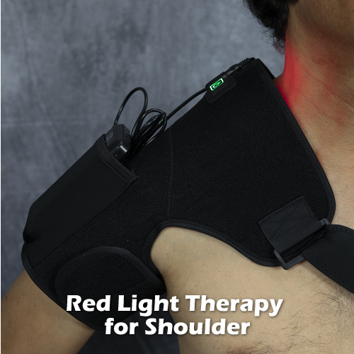
You can use a red light therapy wrap to reduce pain and swelling of your impingement syndrome. Red light therapy wraps use light in the therapeutic wavelengths of red and infrared to accelerate tissue healing and deep heat tissues to increase blood flow and oxygen. See below for an example:
SUMMARY:
Shoulder impingement syndrome occurs when a swollen tendon and/or bursa inside the glenohumeral joint, where your humerus articulates with your scapula, impedes movement of the joint by rubbing against hard structures, particularly the acromion. The goal is to shrink the swollen tendon so that proper movement is restored, and then correct any biomechanical deficiencies in the shoulder complex, such as weak or tight muscles, and subluxated joints affecting the shoulder movements, using exercise rehabilitation, joint mobilization and red light therapy.
Last week, I wrote an article about the Three Paradigms of Health Care. Just to clarify, paradigm in this sense means “a typical example or pattern of perceiving or doing something,” such as society’s paradigm of the “ideal” family.
A paradigm doesn’t carry implications of right or wrong; it simply describes how things are currently done or viewed by society. Paradigms materialize from a combination of historical events, new scientific findings and social trends.
So, the paradigm of health care refers to what typical individuals think and expect of health care.
My proposal is that there are three paradigms of health care:
1. Passive healthcare where you receive it from a provider (doctor, therapist), which includes drug prescriptions, surgery, and physical therapy and chiropractic treatment.
2. Doctor-prescribed lifestyle modification directives; i.e. diet restrictions, exercises, work limitations, etc.
3. “Do It Yourself” health care, where the individual researches health information without the help of a doctor and does things to improve his or her health.
My conclusion is that all three are needed to cover all bases.
But, I also insist that the vast majority of people in the world have a sort of mental attachment; almost an addiction to the first paradigm.
There is this ingrained belief that one must have something done to them by a doctor to get healthy. I think it’s mostly due to the barrage of drug advertisements, medical validation by society’s major institutions, and influence from parents.
Not enough emphasis is placed on the idea of nurturing the body and optimizing its natural recuperative abilities through lifestyle modification. As a result, we live in a society that has a reactive stance to health that actually embraces “sick” care, not health care.
We are not as discerning as we should be in what we eat and fail to make time in our schedules to exercise or engage in physical activities like going on a long hike. This makes us vulnerable to disease, illness and pain as it weakens our bodies’ ability to handle its environment. It cracks the door open for cancer, diabetes, heart disease, arthritis, and chronic joint pain to creep into our lives.
Here’s my advice:
If you think you might be giving too much credit or deference to the first paradigm, shift gears in your thinking and investigate the third paradigm, “Do It Yourself” health.
Here are the advantages:
1. When you invest in your own health, i.e. take more responsibility and play an active role in it, you will be healthier. It’s like a person diligently studying for an exam vs. one who doesn’t and tries to wing it on exam day: the guy who is passive will do worse every time. Like the guy who didn’t study, having a passive attitude towards your health leaves you vulnerable and unprepared.
2. For many types of cases, it’s cheaper than going to the doctor or therapist. You can find home treatment solutions to many common ailments online such as back pain, skin rashes, tension headaches and so on. The key is to read only reputable sites (do research on the individual providing the information first to confirm he/she is qualified).
I’m not saying to never visit your doctor; just pointing out that in this day and age, information on anything is highly accessible and should be utilized properly. Now I know there is a lot of crap online as well; the key is to develop an eye for what is legitimate and what is not, and do your due diligence.
So the question I present is, “Is it possible to cure pain by yourself?”
The answer is YES. Now, that is just an answer to the question from a logic standpoint. I’m not saying that it is possible for you specifically; just that it is possible.
The pain I am talking about is chronic musculoskeletal pain, or pain affecting the bones, joints, muscles and related connective tissue (ligaments, tendons, cartilage).
Obviously there is a “point of no return” where these structures are so degenerated or damaged that pain will always be present. This applies to conditions like knee, hip and low back pain involving severe, irreversible degeneration of structures. What’s critical, then, is that you take action BEFORE you reach that point of no return; and there are LOTS of things you can do to arrest or even reverse the progression of degeneration.
The biggest one is dietary changes. If you have some kind of chronic pain, have you considered using dietary intervention to stop the pain?
Dietary intervention is a popular means to achieve weight loss, but you rarely hear about it being used specifically to fight pain.
Eliminating certain food categories can go a long way in reducing pain. This includes processed sugar, grains, preservatives, additives, alcohol and caffeine. Try doing this for just one week, and see how you feel. I think you’ll be amazed.
Conversely, adding certain food categories or increasing their intake can reduce pain. This includes deep green, leafy vegetables; restricting fat intake to healthy fats like egg yolks, olives, coconut, avocados, seeds and nuts; and drinking only water. Do this for just one week, and see how feel. I think you’ll be amazed.
Secondly, there are home therapies you can do to reduce pain. Massage, joint mobilization and certain safe, therapeutic tools and equipment are available. One of my favorites that I use myself (for prevention) and have prescribed to patients suffering from low back pain is the PosturePump.
Lastly, making some tweaks to your routine can go a long way in reducing and reversing pain. This includes restful sleep, grounding, standing more and sitting less, and doing mind-body techniques such as meditation and EFT.
Bottom line, the Third Paradigm of health care, Do It Yourself health care is on the rise. There is still a cautious attitude towards it within the medical field, but probably because the idea of “people doing what doctors are trained to do” doesn’t sit well with many doctors. But don’t let this discourage you. Remember, you have the biggest stake in your health, so it is prudent to be invested in your health. Don’t simply delegate it to doctors; get yourself involved; know what’s going on, what’s available for treatment, and the risks.
Receive a FREE, 30-Day Plan to Boost Your Health and Eliminate Pain!
As a subscriber, you'll also learn the special methods used by experts in human biomechanics to fix body aches and pain the RIGHT way, long term.
We'll also send you a Free eBook, Concepts of Self-Healing as a way of saying thanks.
Please check your email in 5 minutes to access your Special Report. Make sure to whitelist "[email protected]" in your email client (Gmail, Yahoo, Outlook, etc.) so that you don't miss this valuable information. One way is to add this email to your email Contacts.


 Anatomy of the Shoulder
 Signs and Symptoms of Shoulder Impingement Syndrome
 Imaging Studies
 Last week, I