What is Stiff Person Syndrome?
The singer Celine Dion, whose glorious singing career spanned the last four decades, was recently diagnosed with a condition called Stiff Person Syndrome. Unfortunately, the condition is prevening her from performing, due to its debilitating effect on muscle control, including the vocal cord muscles.
Stiff Person Syndrome (SPS) is a rare, progressive neurological disorder characterized by significant muscle rigidity and spasms, often leading to debilitating physical and psychological symptoms. This article provides an overview of SPS, delving into its causes, symptoms, diagnosis, and current treatment options, as informed by medical research and trusted medical resources.
What is Stiff Person Syndrome?
SPS is an autoimmune neurological disorder primarily causing muscle stiffness and painful spasms. These spasms can worsen over time and vary in symptoms, including an unsteady gait, double vision, or slurred speech, depending on the SPS type. The condition can be severely disabling, often leading to hunched over postures and frequent falls due to impaired reflexes.
Causes of Stiff Person Syndrome
While the exact cause of SPS is unknown, it is believed to be an autoimmune reaction where the immune system attacks glutamic acid decarboxylase (GAD) in the brain and spinal cord. GAD is crucial for producing gamma-aminobutyric acid (GABA), a neurotransmitter controlling muscle movement. Neurotransmitters are protein molecules released from the ends of neurons, which then attach to other neurons causing them to continue the nerve impulse until it reaches the muscle. Disruption in GABA production can lead to continuous neuron firing, contributing to muscle rigidity and spasms seen in SPS. Low GABA levels are also associated with anxiety and depression. Interestingly, SPS often occurs alongside other autoimmune diseases like type-I diabetes, thyroiditis, vitiligo, and pernicious anemia.
Symptoms of Stiff Person Syndrome
The primary symptoms of SPS include progressive muscle rigidity and painful spasms, often triggered by stimuli such as noise, touch, and emotional distress. Initial symptoms typically manifest between the ages of 30 and 60 and can vary in severity and progression. Common initial signs include muscle stiffness and pain, especially in the lower back and legs, potentially leading to difficulty in walking and performing daily activities. Severe cases may require wheelchair use, and there’s an increased risk of anxiety and depression.
Diagnosis of Stiff Person Syndrome
Diagnosing SPS is challenging due to its rarity and symptom overlap with other conditions like Parkinson’s disease, multiple sclerosis, and fibromyalgia. A definitive diagnosis is often made via a blood test measuring GAD antibodies. Most people with SPS show elevated GAD antibody levels. Electromyography (EMG) tests can also be employed to measure muscle electrical activity and assist in diagnosis and monitoring treatment response.
Treatment Options for Stiff Person Syndrome
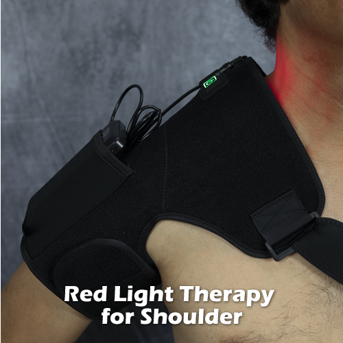
While there is no cure for SPS, symptoms can be managed through personalized treatment plans focusing on pain management, muscle relaxation, and immune response suppression. Common medications include pain relievers, muscle relaxants, anti-seizure and anti-anxiety drugs, sedatives, and steroids. Non-medication treatments like physical therapy, heat therapy, red light therapy, and pulsed EMF are also beneficial. In cases where medications are ineffective, treatments like Botox, intravenous immunoglobulin (IVIg), and stem cell therapy have shown promise in symptom improvement.
Additionally, a narrative review of available medication treatments for SPS suggests starting with benzodiazepines as a first-line treatment, adding medications like levetiracetam or pregabalin if symptoms persist. For second-line therapy, oral baclofen is preferred over rituximab and tacrolimus. In cases of refractory symptoms, treatments like intrathecal baclofen, IVIG, or plasmapheresis can be effective, with intrathecal baclofen and IVIG being more effective than plasmapheresis.
Conclusion
Stiff Person Syndrome presents a complex clinical challenge due to its rarity, varied symptomatology, and the intricate interplay of autoimmune responses. Understanding its underlying causes, symptom patterns, and current treatment modalities is crucial for effective management. Ongoing research continues to shed light on this condition, offering hope for more effective treatments in the future. For individuals diagnosed with SPS, a collaborative approach involving neurologists, rheumatologists, and physical therapists, alongside personalized treatment strategies, is key to managing this condition and improving quality of life.
And lastly, when your body is struggling with disease, give it assistance by providing it with nutrients, water, sunlight and mild exercise when possible. Mind-body approaches including meditation, flotation therapy, biofeedback, yoga, tai-chi, deep breathing and so on, may provide some relief as well.
Sources:
Johns Hopkins Online
https://www.hopkinsmedicine.org/
National Institute of Neuromuscular Disorders and Stroke
American Brain Foundation


 The quality of the air we breathe can have significant effects on our health, particularly when it contains fine particulate matter (abbreviated “PM2.5”). Understanding the consequences of inhaling PM2.5 can underscore the importance of maintaining air quality and safeguarding our respiratory health.
 In late summer of 2020, the Bay Area of California experienced very dangerous levels of PM2.5 due to wildfires; so bad that people were instructed to remain indoors. And on the day of and weeks following the 9-11 terrorist attacks on the World Trade Center, thousands of people, especially first responders, breathed in the tons of fine particulate matter that were produced from the destruction. Many are
 Could This be the Best Home Air Purifier Ever Made
 Anatomy of the Shoulder
 Signs and Symptoms of Shoulder Impingement Syndrome
 Imaging Studies